This article has been reviewed according to Science X's editorial process and policies. Editors have highlighted the following attributes while ensuring the content's credibility:
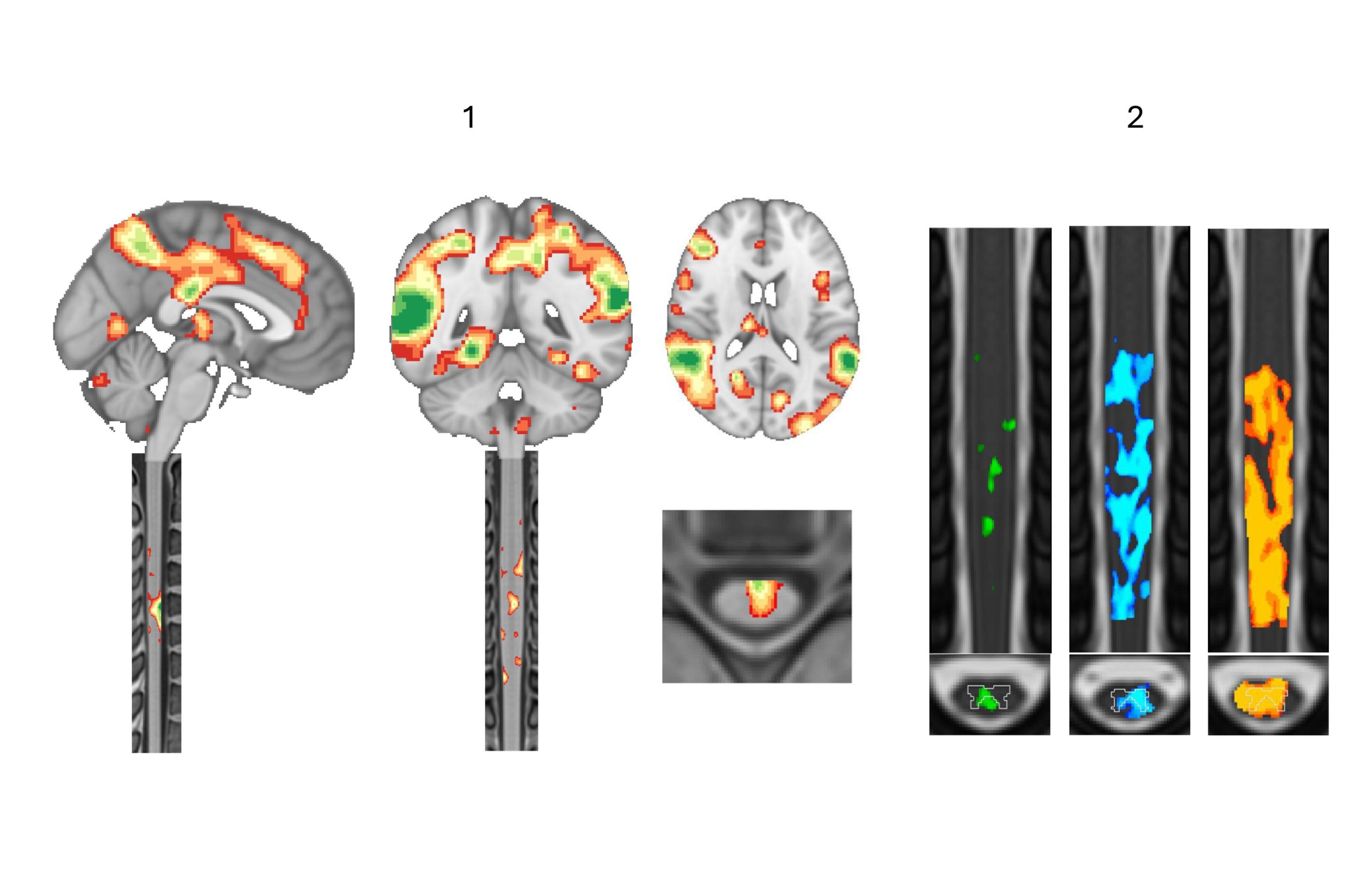
Several EPFL labs are converging around an idea that began as a doctoral researcher's hunch: The spinal cord revealed by functional magnetic resonance imaging might unlock new therapies.
"Have you seen Galileo's drawings of the moon?" I ask Nawal Kinany, a former EPFL researcher who now works in international scientific cooperation. She hasn't, so I pull them up on my laptop. In 1610, working from a blurry telescope, Galileo employed his talents as a draftsman to infer craters and mountains where others had only seen blotches. Kinany spent eight years on a different blur: the faint, noisy signal that functional magnetic resonance imaging, or fMRI, picks up from the human spinal cord. The picture she helped sharpen now guides treatments for paralysis and may soon do the same for stroke and Parkinson's.
Functional magnetic resonance imaging, a brain imaging technology, records neural activity without actually seeing the neurons fire. It tracks the rush of oxygen-rich blood that follows them, a few seconds late, one snapshot every two or three seconds. Stitched together, the snapshots show not only where activity happens but how it shifts. Ask a woman lying in the scanner to tap her fingers, and a patch of her brain lights up; when she stops, it fades. The same tap lights up her spinal cord too , just below the neck, although that signal is far harder to catch.
Source link