This article has been reviewed according to Science X's editorial process and policies. Editors have highlighted the following attributes while ensuring the content's credibility:
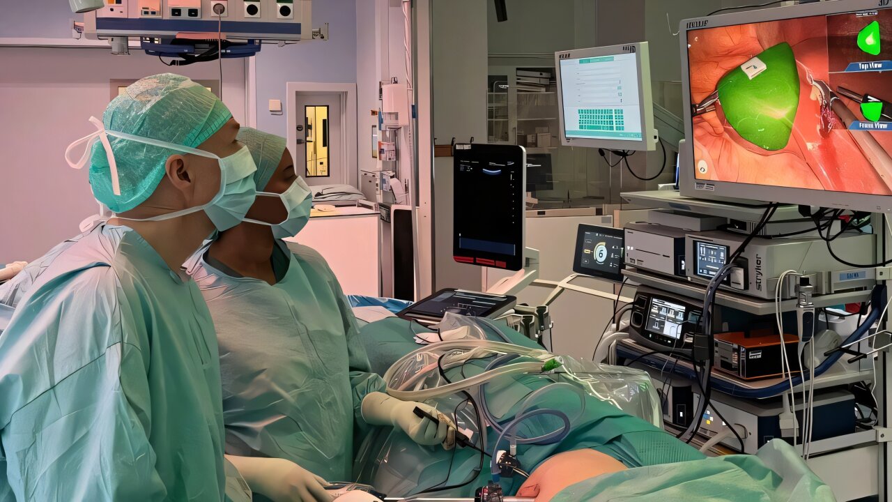
Tumors beneath the surface of the liver can be difficult to locate during laparoscopic, or keyhole, surgery. The new technology uses augmented reality to overlay a 3D visualization of the tumor and other anatomical structures on the surgeon's camera view. The visualization is updated as the camera changes position.
"We have brought together the geometric tools needed to show where the tumor would be visible if the liver were semitransparent," says Torbjörn Lundh, professor at the Department of Mathematical Sciences at Chalmers University of Technology and the University of Gothenburg.
"You can think of it as a GPS inside the organ. Instead of just having a map before surgery, we get continuous guidance based on the current camera angle," says Niclas Kvarnström, transplant surgeon and head of the Transplant Centre at Sahlgrenska University Hospital, and a researcher at Sahlgrenska Academy at the University of Gothenburg.
In June 2026, the technology was used in surgery on a patient for the first time.
"The system could be used as planned during the operation, and the workflow worked well. The patient was able to go home the following day," says Kvarnström.
To use the technology, the surgeon places a special sticker on the surface of the liver. 16 square inches) in size and has a QR code. As long as the camera can see the marker, the system can show the tumor or other structures beneath the surface.
Source link