This article has been reviewed according to Science X's editorial process and policies. Editors have highlighted the following attributes while ensuring the content's credibility:
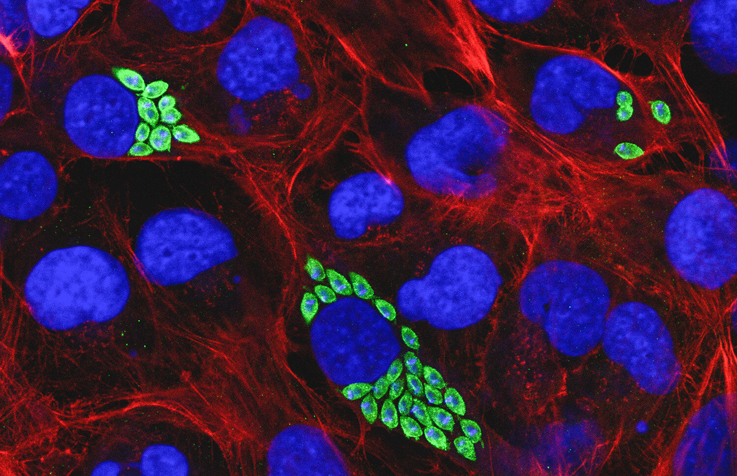
By combining live imaging of brain tissue with artificial intelligence, researchers at Uppsala University have developed a method that reveals how individual glioblastoma cells spread through the brain, how they can switch between different ways of moving and how they are affected by cancer treatments. An increased understanding of tumor cell invasion might in the long term contribute to more effective therapies.
Glioblastoma is one of the most aggressive forms of brain cancer. One reason it is so difficult to treat is that tumor cells spread into the surrounding healthy brain, beyond the area that can be removed by surgery. These remaining cells can contribute to the cancer returning after treatment.
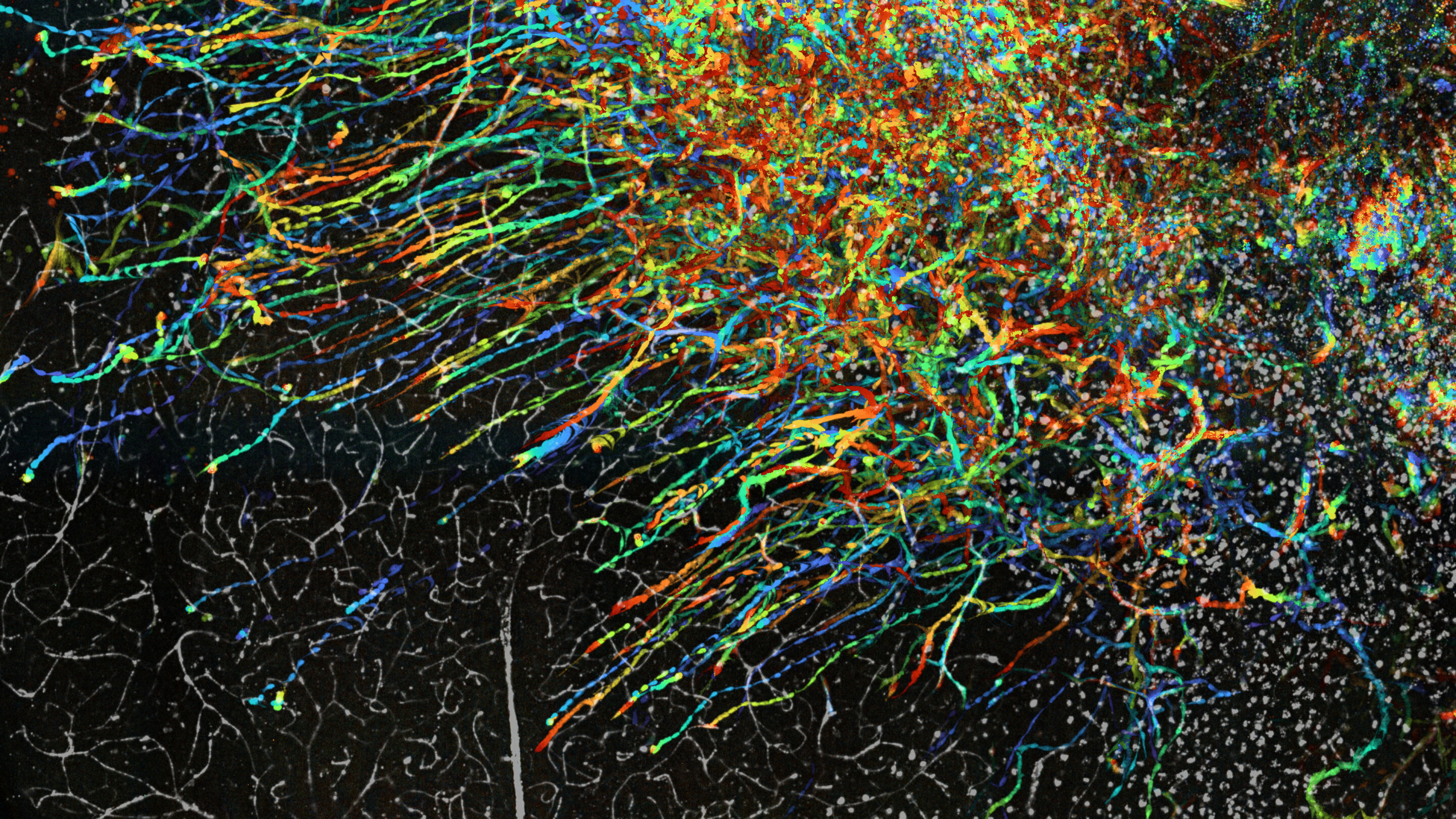
In a study in the journal Nature Communications, researchers at Uppsala University present GlioTrace, a new method that can follow thousands of individual tumor cells as they move through brain tissue.
"Until now, we have mostly seen snapshots of how glioblastoma spreads. This method combines microscopy with artificial intelligence and allows us to watch the process unfold in living brain tissue. We could also compare tumors from different patients and investigate how they respond to treatment," says Hitesh Mangukiya, a researcher at the Department of Immunology, Genetics and Pathology at Uppsala University and one of the study's lead authors.
The researchers studied glioblastoma cells originating from six patients. The cells were grown as tumors in mouse brains, which were then cut into thin slices and kept alive in the laboratory.
Source link