This article has been reviewed according to Science X's editorial process and policies. Editors have highlighted the following attributes while ensuring the content's credibility:
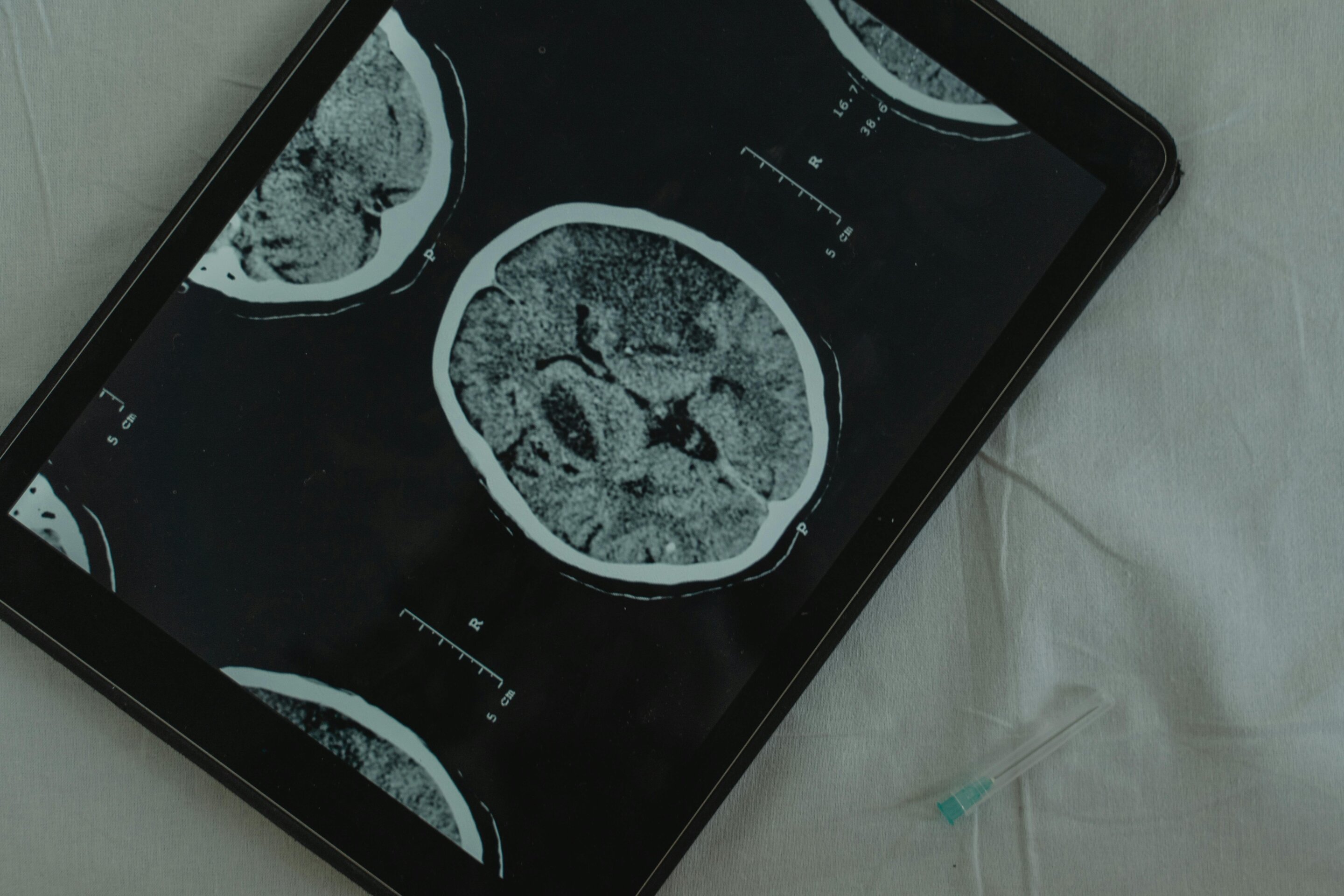
In 2007, researchers in the Netherlands scanned the brains of 2,000 people aged 45 and over who were going about their ordinary lives. Seven in every 100 had signs of a stroke they never knew they'd had. Nearly two in 100 had a small bulge in a blood vessel, and a similar number had a tumor that appeared to be benign, meaning it was not cancerous.
Yet only two of those 2,000 people reported symptoms that could be linked to what the brain scanner found. Both had hearing loss.
That finding sits at the heart of how brain scans are used in medicine. A scan shows what the brain looks like but it cannot, on its own, tell you why someone is unwell. Joining those two things up is the hard part, and it is still mostly done by people rather than machines.
The most detailed pictures of the brain's structure often come from MRI, which uses a powerful magnet and radio waves rather than X-rays. It can reveal a tumor, bleeding, the scar left by a stroke or head injury, or a patch of brain that didn't develop quite as it should have before birth. Some of these changes are smaller than a grain of rice.
What a standard MRI can't show is how the brain's electrical activity changes from moment to moment.
Source link