This article has been reviewed according to Science X's editorial process and policies. Editors have highlighted the following attributes while ensuring the content's credibility:
An international team of scientists has discovered how some of the tiniest structures in the human spine help it cope with the strain of bending, lifting and everyday movement, potentially shedding new light on why spinal disks deteriorate.
The study is published in Nature on Nov. 4 and involves researchers at The University of Manchester, Queen Mary University and University College London, in collaboration with the ESRF—the European Synchrotron—and Diamond Light Source.
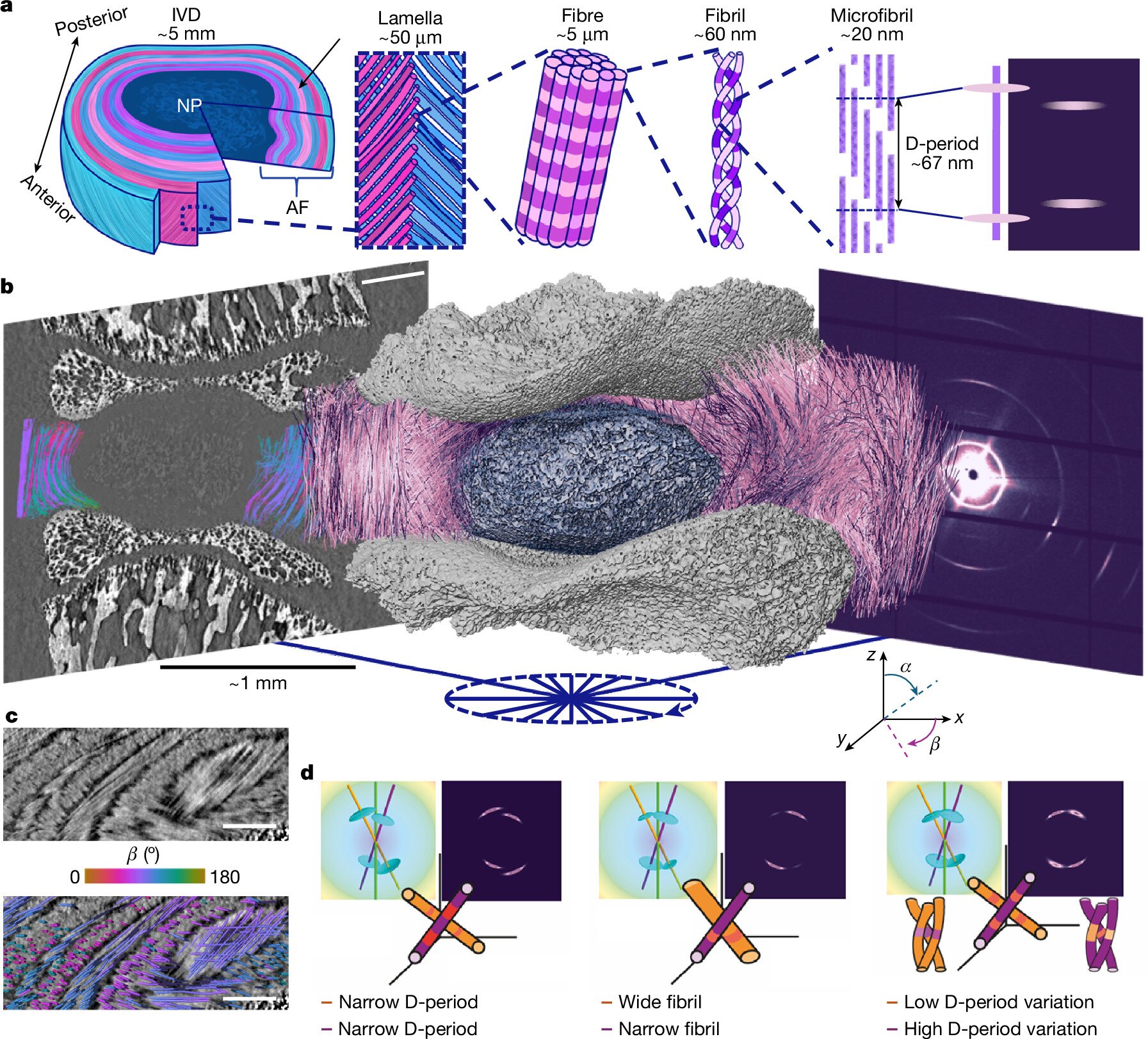
They used powerful X-rays to examine what happens inside intervertebral disks at scales ranging from whole tissue down to individual collagen structures.
The imaging method, called TomoSAXS, combines different X-ray techniques to reveal in 3D how the collagen fibers that give disks strength respond when compressed.
They found collagen fibrils—tiny, strong strands made from many collagen molecules packed tightly together—are already under strain before additional pressure is applied, which may help disks withstand everyday loads but could also make some structures more vulnerable to damage.
Highly curved collagen fibers experienced particularly high strain, especially around the boundary between the disk's inner and outer regions, an area that may be more susceptible to damage and herniation.
Dr. Alissa Parmenter, joint first author, from University College London, said, "When we bend or lift, the forces within our spinal disks are shared between structures far smaller than we can see. With TomoSAXS, we can follow how the tissue deforms and connect that movement to changes within its nanoscale collagen building blocks.
Source link