This article has been reviewed according to Science X's editorial process and policies. Editors have highlighted the following attributes while ensuring the content's credibility:
The brain's motor cortex plans, controls and executes voluntary muscle movements. Whether you're playing catch, writing your name or chewing food, it all points back to this region and its connections to other parts of the brain. Yet despite extensive study, scientists still haven't created a detailed map of how the mouse motor cortex is internally organized. Most brain atlases have divided it into two general regions—primary and secondary—based on differences in connectivity, but this doesn't provide a detailed or consistent picture.
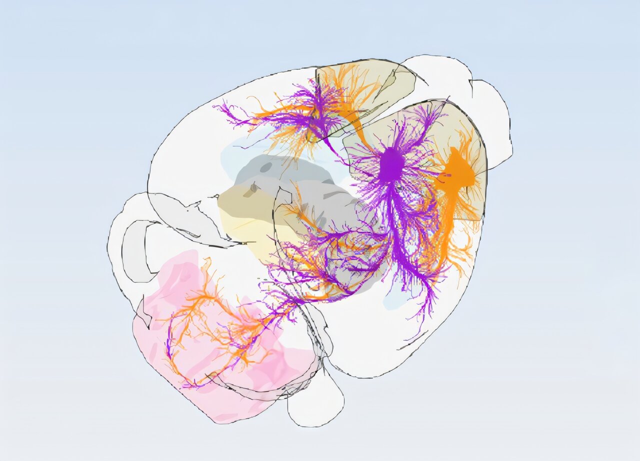
A new study published in Cell by researchers at the University of Basel, Friedrich Miescher Institute for Biomedical Research and the Allen Institute paints a more precise portrait of the mouse motor cortex. Scientists mapped this area into 16 distinct subregions, each with its own pattern of connections. Their results could have important implications for understanding diseases that affect the motor cortex, such as amyotrophic lateral sclerosis (ALS) and frontotemporal dementia (FTD).
"Understanding the precise wiring of the brain is essential for developing effective treatments for brain diseases. Here, our Swiss collaborators' expert charting of the functionally specific motor cortex circuit, combined with the Allen Institute's foundational connectivity atlas, resulted in such a precision map that drives movement control," says Hongkui Zeng, executive vice president and director of Brain Science at the Allen Institute and a senior author on the study.
Researchers studied 547 experiments that tracked how nerve signals travel out of the motor cortex and into other parts of the brain.
Source link