This article has been reviewed according to Science X's editorial process and policies. Editors have highlighted the following attributes while ensuring the content's credibility:
The brain is often described in terms of cells and wiring. However, much of it is made of lipids that form the membranes of neurons, wrap nerve fibers with myelin and help brain cells send and receive signals.
Despite their importance, lipids have been a blind spot in neuroscience. Unlike genes and proteins, which have been mapped in detail, lipid molecules are difficult to distinguish with standard imaging methods.
"So scientists have had only a blurry picture of how lipids differ from one part of the brain to another," says Giovanni D'Angelo, professor at EPFL's Kristian Gerhard Jebsen Foundation Chair in Nutrition and Metabolism. "That gap matters, because changes in brain lipids are increasingly linked to conditions from depression to Alzheimer's disease."
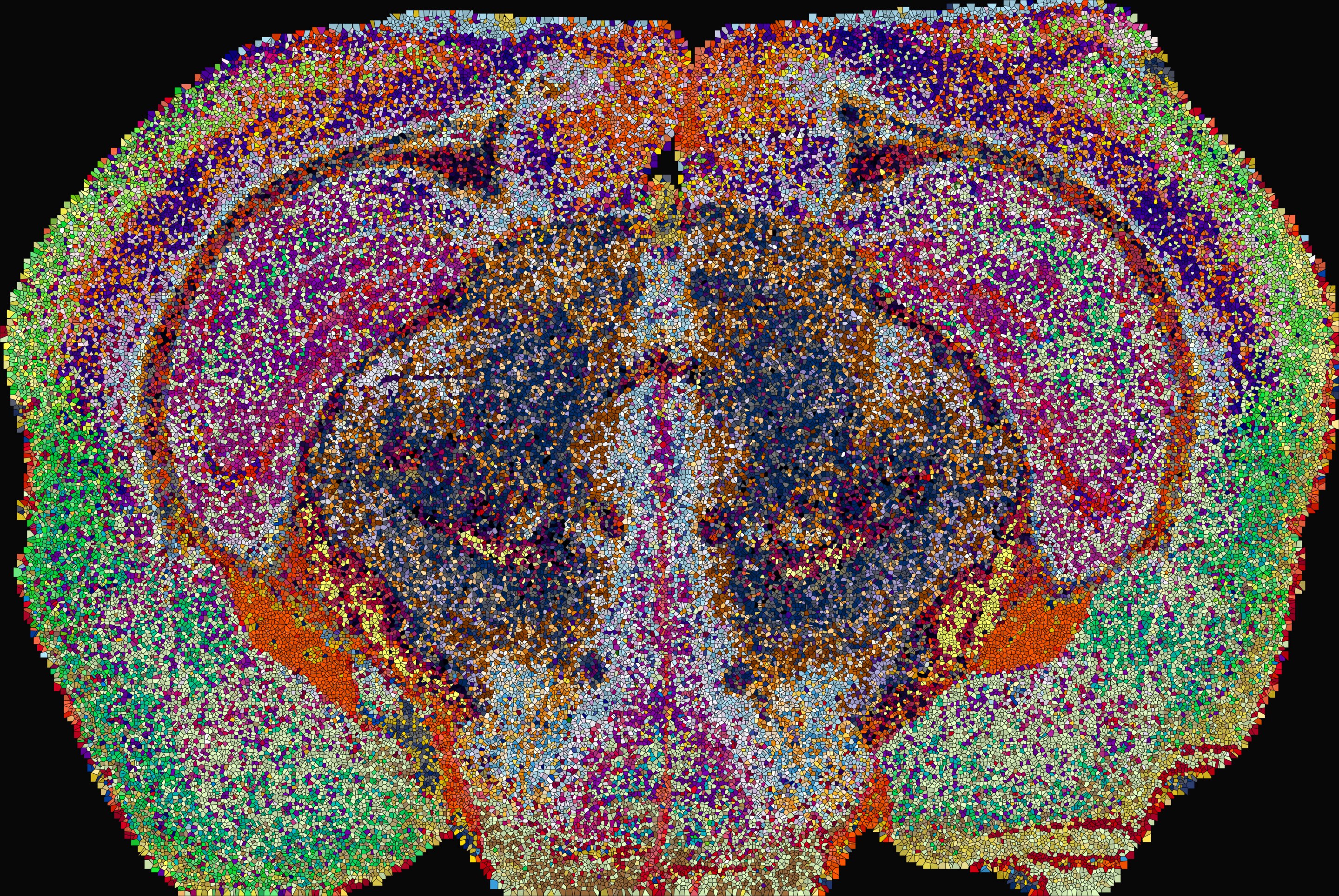
Now, a team of scientists led by D'Angelo, Luca Fusar Bassini and Gioele La Manno at EPFL has built a detailed atlas of lipids across the entire mouse brain. The Lipid Brain Atlas shows that these molecules follow a precise, orderly pattern that lines up with the brain's known anatomy and reveals boundaries that anatomy alone has missed. The pattern is so distinctive that, from the lipids alone, the team could tell where in the brain a piece of tissue came from.
To build the atlas, the team used a method called mass spectrometry imaging. It fires a laser at thin slices of brain, point by point, and records which lipids sit at each spot.
Source link