This article has been reviewed according to Science X's editorial process and policies. Editors have highlighted the following attributes while ensuring the content's credibility:
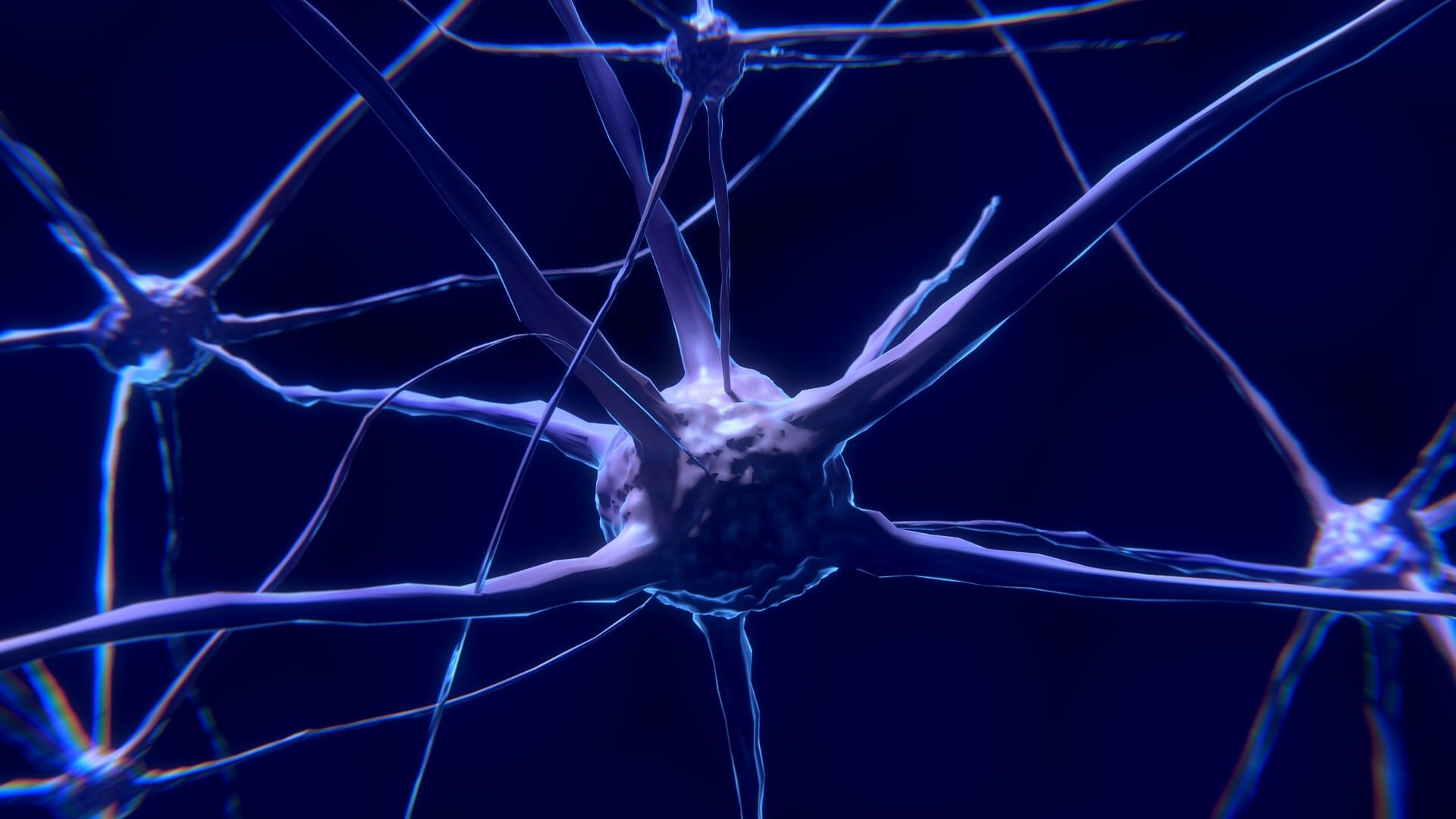
Scientists studying amyotrophic lateral sclerosis (ALS), Parkinson's, Alzheimer's and other neurological diseases are getting unprecedented views inside brain cells, using powerful new microscopes to uncover changes that may begin long before neurons die. These technologies enable researchers to zoom in on individual neurons and the tiny structures inside them, watch cells at work in real time and use artificial intelligence to identify patterns linked to disease, according to experts who spoke at the Oct. 18 Presidential Symposium—"Advances in Microscopy for Visualizing Neurologic Disease"—at the 151st Annual Meeting of the American Neurological Association (ANA 2026).
"Neurological diseases ultimately arise from changes in cells and the extraordinarily complex structures within them. Until recently, many of those changes could not be visualized," said Dimitri Krainc, M.D., Ph.D., FANA, president of the ANA, chair of the symposium, chair of the Davee Department of Neurology, director of the Feinberg Neuroscience Institute and Aaron Montgomery Ward Professor at Northwestern University Feinberg School of Medicine in Chicago.
"These technologies are giving us a much more detailed and dynamic view of disease biology, with the potential to reveal abnormalities earlier and point us toward treatments that address the underlying molecular disruptions in neurons."
, FANA, is examining the endoplasmic reticulum (ER), an organelle network inside cells that performs several essential jobs. Advanced microscopy approaches reveal that parts once thought to be flat sheets are constantly changing networks of tiny tubes, providing a new view of how cells work and what may go wrong in diseases linked to mutations in proteins, including inherited neurological disorders such as hereditary spastic paraplegias.
Source link