This article has been reviewed according to Science X's editorial process and policies. Editors have highlighted the following attributes while ensuring the content's credibility:
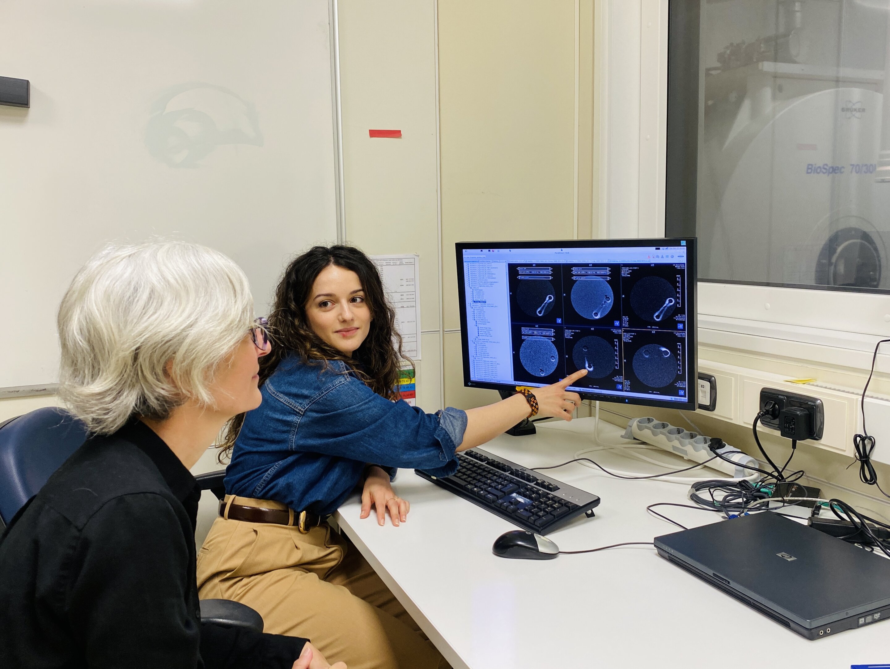
The Biomolecular Nanotechnology research group led by Ikerbasque research professor Aitziber L. Cortajarena has developed a new iron-based contrast agent for magnetic resonance imaging that can be used for both diagnosis and treatment. In a study carried out on lab-grown human heart tissue, it was proven that the contrast agent accumulates in the fibrotic tissue, that its signal intensity on the MRI image increases with the severity of the disease, and that it slows disease progression.
The study, led by Cortajarena and conducted in collaboration with Ana Villar's group at the Institute of Biomedicine and Biotechnology of Cantabria (IBBTEC, UC-CSIC), has been published in the journal Advanced Functional Materials. It paves the way for a possible alternative to gadolinium, the metal currently used in almost all contrast-enhanced MRI scans.
Cardiac fibrosis is one of the main factors leading to heart failure, a disease that affects millions of people worldwide. There is currently no approved treatment specifically designed to slow down this condition. When the heart is damaged or under sustained strain (for example, after a heart attack or due to hypertension), a repair process begins, similar to the healing of a cut on the skin.
The collagen production process is activated to repair and strengthen the tissue. However, this process often does not stop: the collagen continues to accumulate, and the heart becomes stiff and finds it increasingly difficult to fill and pump blood properly.
Source link