This article has been reviewed according to Science X's editorial process and policies. Editors have highlighted the following attributes while ensuring the content's credibility:
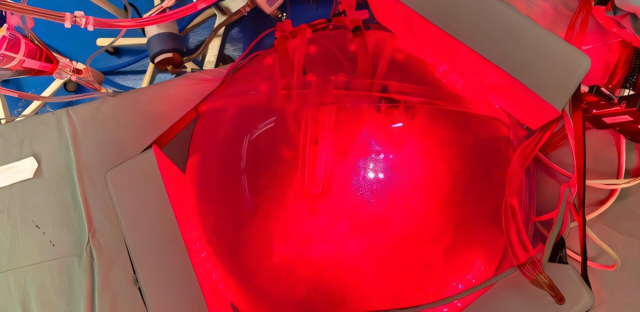
Researchers at the University of São Paulo (USP) in Brazil and their international collaborators have created a computational model that can generate a "digital twin" of the human thorax. The goal is to personalize the treatment of respiratory diseases using a special type of light.
Described in a paper published in PNAS, the new technology uses CT scans to predict the path that light radiation will take inside the body. This makes it possible to calculate the exact dose needed to combat infections and inflammation directly in the lungs by illuminating the person from the outside.
Light-based therapies have been used to treat inflammation or infections in various organs. The two main approaches are photodynamic therapy, which uses light to activate a photosensitive substance that generates reactive species capable of destroying microorganisms in the presence of oxygen, and photobiomodulation, which uses low-intensity light to modulate cellular processes and inflammatory responses.
However, in the case of the lungs, the use of these techniques presents a significant challenge. To reach the organ noninvasively, the externally applied light must pass through the skin, fat, muscles, bones and other chest tissues, undergoing absorption and scattering along the way.
Thus, the same amount of light applied to two people's chests can produce very different effects in their lungs. To address this issue, the authors combined computed tomography images, three-dimensional anatomical reconstructions and computational simulations accelerated by high-performance computing.
Source link