This article has been reviewed according to Science X's editorial process and policies. Editors have highlighted the following attributes while ensuring the content's credibility:
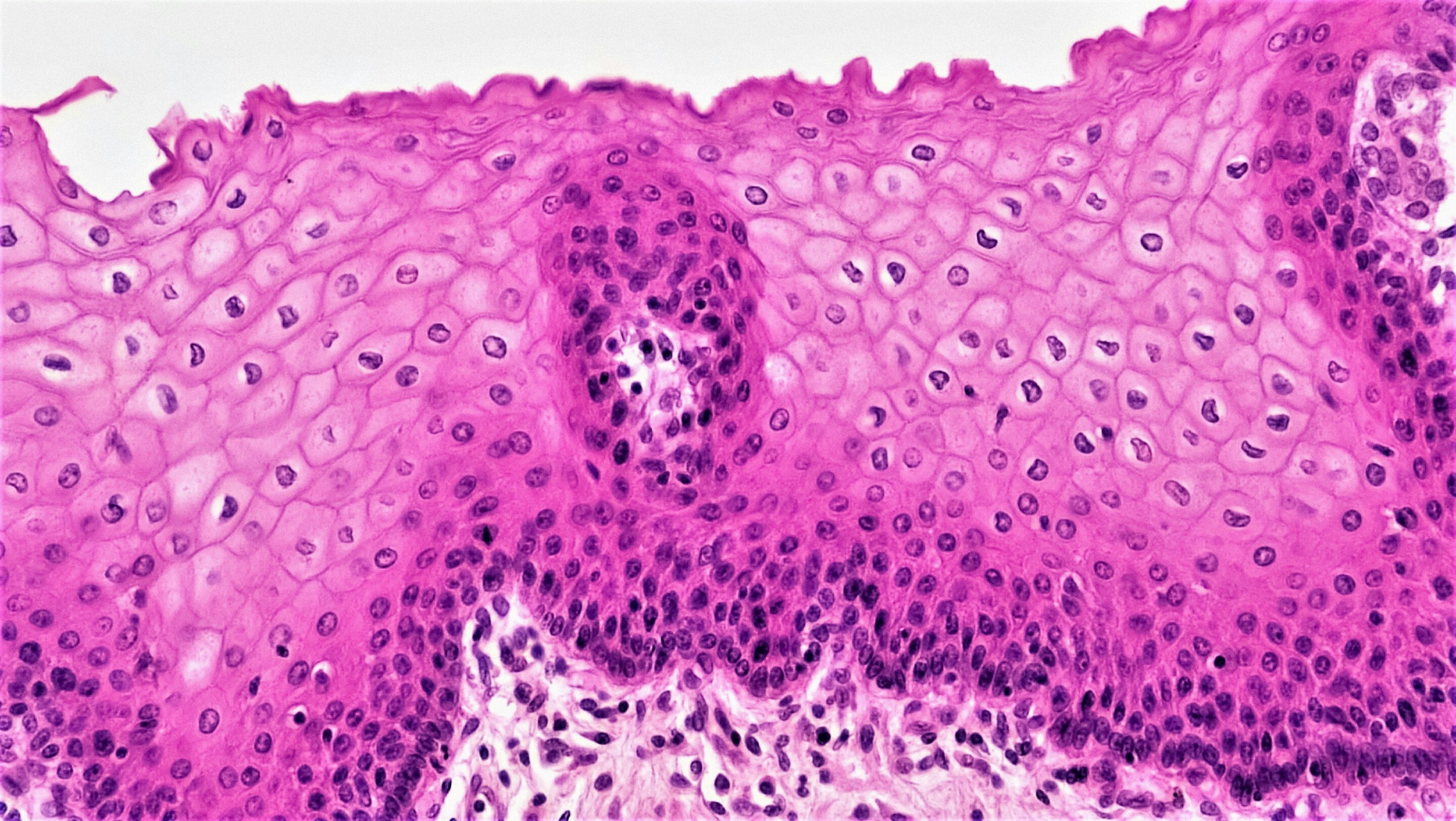
Artificial intelligence can help identify cells in digital pathology images, but densely packed tissue remains a challenge: neighboring cells may be merged, counted twice or missed entirely. Researchers at Koç University have developed a training strategy that improves detection by teaching AI to consider how cells are arranged within the tissue.
Published in Medical Image Analysis, the study introduces topology-guided hard example mining (TG-HEM). The approach helps models concentrate on image regions where their predictions fail to capture the spatial organization of cells, rather than relying only on errors at individual pixels.
The research was conducted by Professor Çiğdem Gündüz Demir, Ph.D. student Onur Çakı and colleagues from Koç University's Department of Computer Engineering and KUIS AI Center, working with collaborators in mathematics and pathology.
Cell counts and spatial distributions provide information that pathologists use when assessing disease and planning treatment. Yet a single tissue image can contain tens of thousands of cells, making manual counting time-consuming and subject to variation between observers.
Deep learning systems can automate this task, but local visual cues can become ambiguous when cells lie close together. The researchers' approach brings information about the surrounding cell arrangement into the training process.
During training, TG-HEM compares predicted cell distributions with annotated reference distributions in sampled image regions. It uses topological data analysis—a mathematical approach for characterizing the structure of data—to quantify differences in their organization.
Source link