This article has been reviewed according to Science X's editorial process and policies. Editors have highlighted the following attributes while ensuring the content's credibility:
A next-generation total-body PET/CT scanner can produce diagnostic-quality images in as little as 1 minute of scanning, according to research presented today at the 39th Annual Congress of the European Association of Nuclear Medicine (EANM'26). Even its fastest images were rated more highly on average than those from a standard PET/CT scanner.
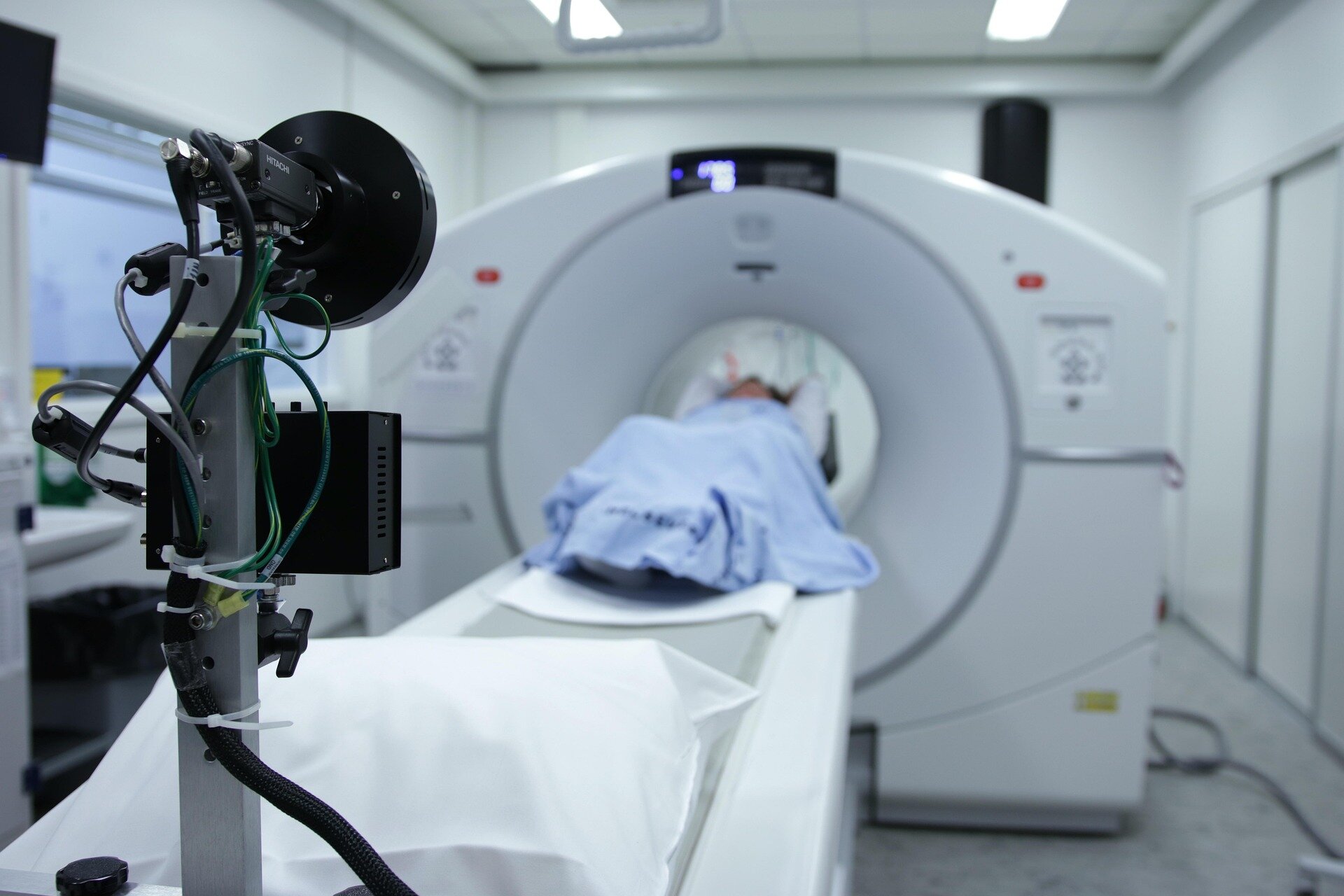
PET/CT is widely used to diagnose, stage and monitor cancer. Conventional PET/CT scanners have a short axial field of view, meaning they image the body section by section, typically requiring five to six 'bed positions' to cover the body from head to thigh. Long-axial field-of-view (LA-FOV) systems capture a much larger part of the body at once and detect more of the signal emitted by the radiotracer, raising the possibility of much shorter scans without sacrificing image quality.
In the EQUIP study, researchers at the Peter MacCallum Cancer Centre in Melbourne, Australia, evaluated a next-generation 128 cm LA-FOV PET/CT system (GE HealthCare Omni 128 cm). Twenty-four participants who had received one of five radiotracers (¹⁸F-FDG, ¹⁸F-DCFPyL, ⁶⁸Ga-DOTATATE, ⁶⁸Ga-PSMA or ¹²⁴I) underwent a standard-of-care scan on a conventional short-axial field-of-view (SA-FOV) PET/CT system, followed by a 10-minute scan on the LA-FOV system. The researchers then reconstructed images from the LA-FOV scan using just 5, 3, 2 and 1 minute of data.
Two nuclear medicine specialists, who were unaware of the acquisition times, graded the images on a five-point scale for overall diagnostic quality, sharpness and lesion conspicuity (how clearly lesions stand out from surrounding tissue) and image noise.
Source link