This article has been reviewed according to Science X's editorial process and policies. Editors have highlighted the following attributes while ensuring the content's credibility:
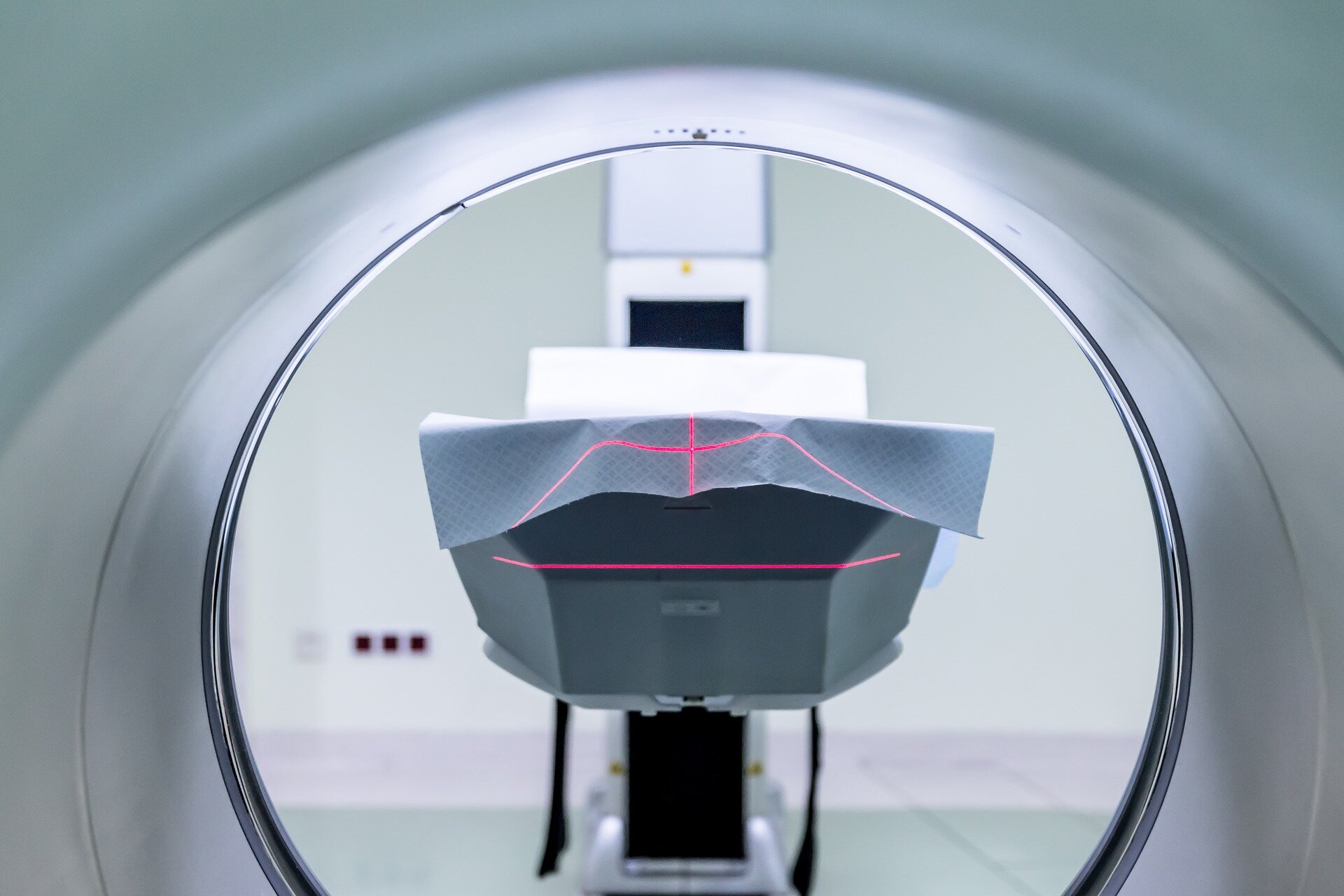
Scientists are developing a new MRI-based test that could spare thousands of cancer patients from aggressive radiotherapy treatment.
The pioneering imaging technology works by "color-coding" oxygen-starved cancerous tissue on scans and could be used to help clinicians identify which patients with HPV-positive throat cancer can receive lower doses of radiotherapy that provide the same treatment benefit while reducing long-term side effects.
The technique, known as HYPOXiMAP, is being developed by researchers at The Institute of Cancer Research, London, in collaboration with University College London and The University of Manchester.
The technique uses advanced MRI to reveal pockets of hypoxia—areas of low oxygen within tumors that are known to make cancer cells more resistant to radiotherapy.
HYPOXiMAP provides this information as a vivid, intuitive color map. Blue areas reveal hypoxic, treatment-resistant tissue, while yellow shows well-oxygenated regions that typically respond well to radiotherapy. Being able to provide this information in real time will one day allow smart radiotherapy systems that can automatically adjust themselves to provide gentler treatment whenever appropriate.
There are around 13,000 new cases of head and neck cancer in the U.K. every year. Rates of HPV-positive oropharyngeal (throat) cancer are increasing sharply, with a 47% rise in cases between 2013 and 2019.
Around 70% of patients are treated with radiotherapy. However, the standard high dose used frequently results in long-term side effects, such as swallowing difficulties, dry mouth, taste changes and persistent fatigue.
Source link