This article has been reviewed according to Science X's editorial process and policies. Editors have highlighted the following attributes while ensuring the content's credibility:
When women develop the hard-to-pronounce lung disease called lymphangioleiomyomatosis (LAM), a natural set of brakes on cell growth fails inside their lungs, causing affected cells to form cysts that can threaten their ability to breathe.
As many as 25% of those who develop LAM die within 10 years, not including those whose lives are extended via difficult-to-obtain lung transplants. In recent years, doctors have used a drug called sirolimus to stabilize lung function, but this treatment is not a cure and is not always well tolerated.
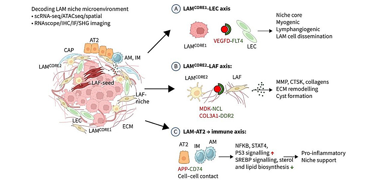
Now a research team led by experts at Cincinnati Children's has produced an extremely detailed "atlas" that details how the various types of cells involved in LAM lose their normal functions and begin interacting in a cancer-like tumor formation process.
"This is the first spatially resolved map of the cells that make up LAM lesions in the lungs of women with this rare disease," says corresponding author Yan Xu, Ph.D., a leading bioinformatics expert and professor in the Perinatal Institute at Cincinnati Children's.
"This map reveals three distinct LAM CORE subtypes with unique signatures, spatial distributions and functions. More importantly, LAM is not just one type of abnormal cell, but a community of cells that communicate with each other," she adds.
Source link