This article has been reviewed according to Science X's editorial process and policies. Editors have highlighted the following attributes while ensuring the content's credibility:
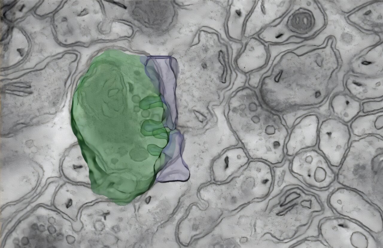
Researchers at Harvard Medical School and the University of California San Diego have sped up a powerful imaging technique enough to map, for the first time, large volumes of brain tissue in nanoscale detail. The approach reveals fine structures that brain wiring maps, known as connectomes, can miss.
The work, described in Nature Methods, promises to help scientists build more complete diagrams of how cells in the brain are connected. This, in turn, could deepen understanding of nervous-system development and certain brain diseases.
"We can now see brain structures that we haven't been able to see before in connectomics data," said co-senior author Wei-Chung Allen Lee, associate professor of neurobiology at HMS and HMS associate professor of neurology at Boston Children's Hospital.
"We hope this work will help us understand how nervous systems are built; what changes with age; and what goes awry in neurodevelopmental, cognitive, and neurodegenerative disorders," Lee said.
Neuroscientists build connectomes to understand how neurons work together to produce thought and behavior.
Some research efforts chart the brain's major highways using MRI scans. Others zoom in, using electron microscopy to trace individual neurons and the synapses where they meet, as in a recently completed connectome of the fruit fly brain and nerve cord that Lee co-led with researchers from around the world.
However, even these fine-grained maps give up some detail when imaging large amounts of tissue.
Source link