This article has been reviewed according to Science X's editorial process and policies. Editors have highlighted the following attributes while ensuring the content's credibility:
The brain's network of blood vessels plays a critical role in maintaining neurological health. Changes to these vessels are associated with conditions including stroke, cognitive impairment and dementia. Yet studying the brain's smallest arteries can be challenging, in part because extracting detailed measurements from MRI images often requires substantial manual work.
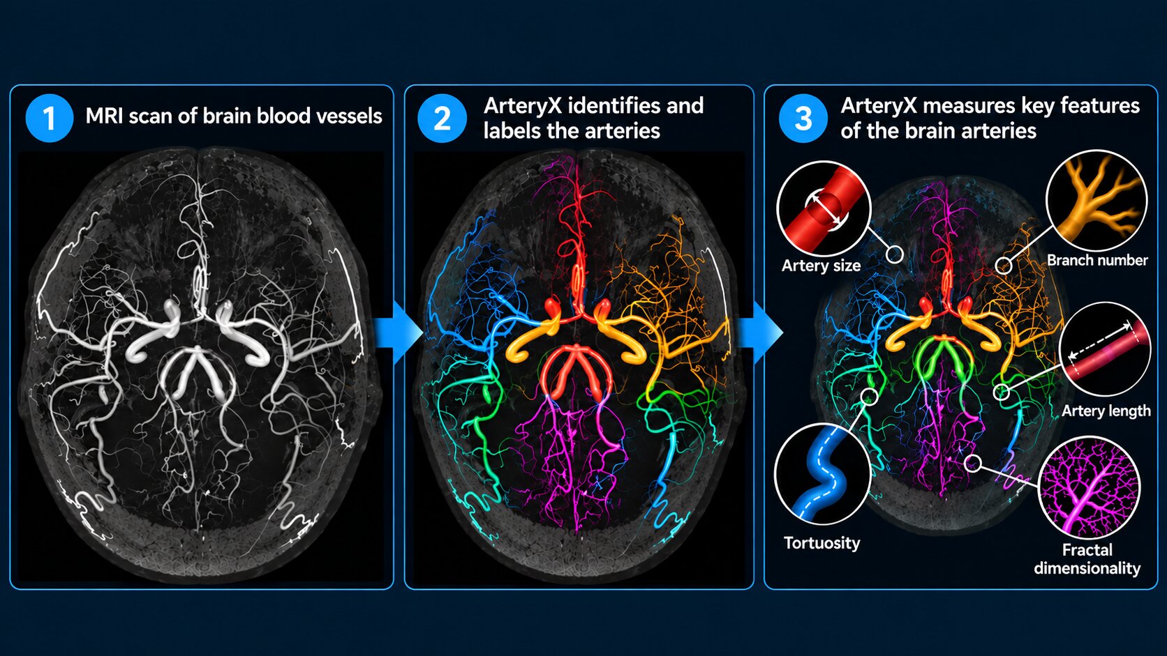
Researchers at University of Rochester Medicine are developing a technology called ArteryX to address this challenge. The software toolbox analyzes MRI images of the brain's arteries, allowing researchers to measure characteristics such as vessel size, length, branching and complexity.
By making this process more automated and consistent, ArteryX could help scientists investigate how changes in the brain's vascular network contribute to disease.
The technology was developed under a project led by Nasir Uddin, Ph.D., and described in a study published in Magnetic Resonance in Medicine. Abrar Faiyaz, Ph.D., a postdoctoral research associate in Uddin's lab, is the study's first author.
MRI can produce detailed images of the brain's blood vessels, but converting those images into standardized measurements has traditionally required researchers to manually trace vessels, correct errors and identify individual arteries. The process is particularly difficult when vessels are small, closely spaced or located farther from the major arteries.
These challenges can make large studies time-consuming and introduce differences between users analyzing the same images. "ArteryX grew out of a major bottleneck in quantitative brain-vessel research," Uddin said.
Source link