This article has been reviewed according to Science X's editorial process and policies. Editors have highlighted the following attributes while ensuring the content's credibility:
In ion beam therapy, atomic nuclei are accelerated to high energies in a particle accelerator, for example, a synchrotron, and precisely directed at a tumor. There, they damage cancer cells and destroy tumor tissue. This technique is now used at several specialized medical facilities around the world, including MedAustron in Wiener Neustadt, Austria.
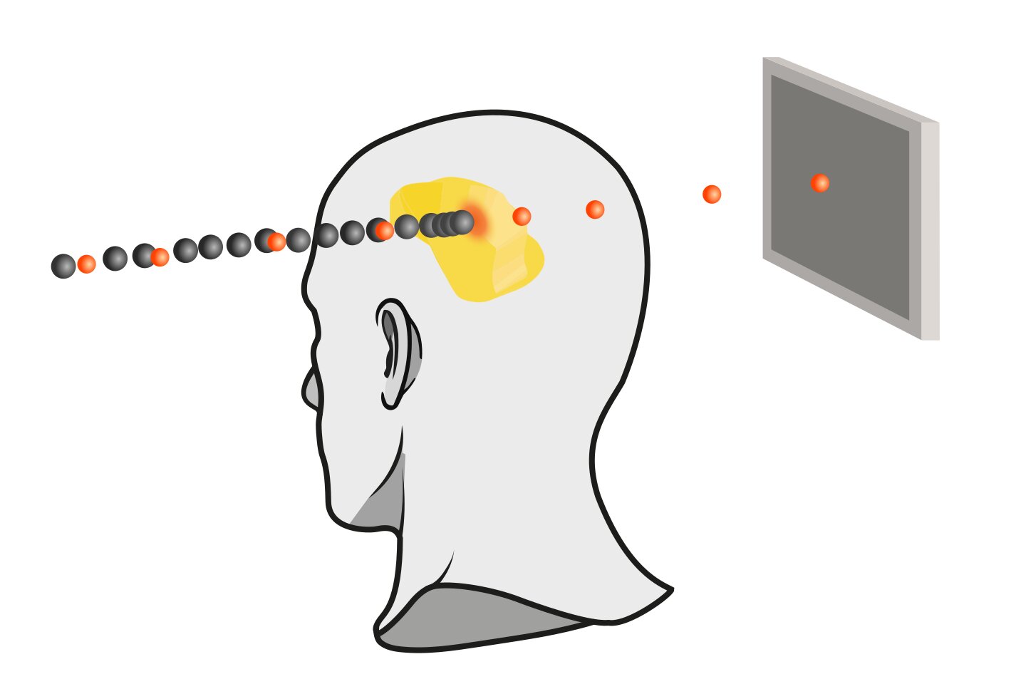
A new approach currently being investigated involves so-called "mixed beams," which consist of two different ion species: Carbon nuclei destroy the tumor cells, while helium nuclei simultaneously help monitor the patient's anatomy during irradiation. To make this possible, however, the precise ratio of carbon to helium in the beam must be known.
But how can an ion beam be measured while it is being used for treatment? TU Wien and MedAustron have now found a solution—an important step on the path toward cancer treatment using mixed ion beams. The paper is published in the journal Physical Review Accelerators and Beams.
Despite the high precision of ion beam therapy, uncertainties remain. Organ motion, for example, can shift the location at which the beam deposits most of its energy—also called the "range" of the beam—by a few millimeters. To ensure that the entire tumor is treated despite these uncertainties, a small safety margin around the tumor is irradiated as well. The smaller this margin can be made, the more precise the therapy becomes and the better sensitive organs nearby can be spared.
Source link