This article has been reviewed according to Science X's editorial process and policies. Editors have highlighted the following attributes while ensuring the content's credibility:
Acute interstitial nephritis (AIN) is a condition characterized by inflammation that damages the kidney's tubules and surrounding tissue. Often resulting from an immune reaction to common medications—including antibiotics, anti-inflammatory medications and drugs that treat acid reflux—the condition accounts for roughly 15% to 20% of biopsied cases of acute kidney injury (AKI), defined as a sudden decline in kidney function.
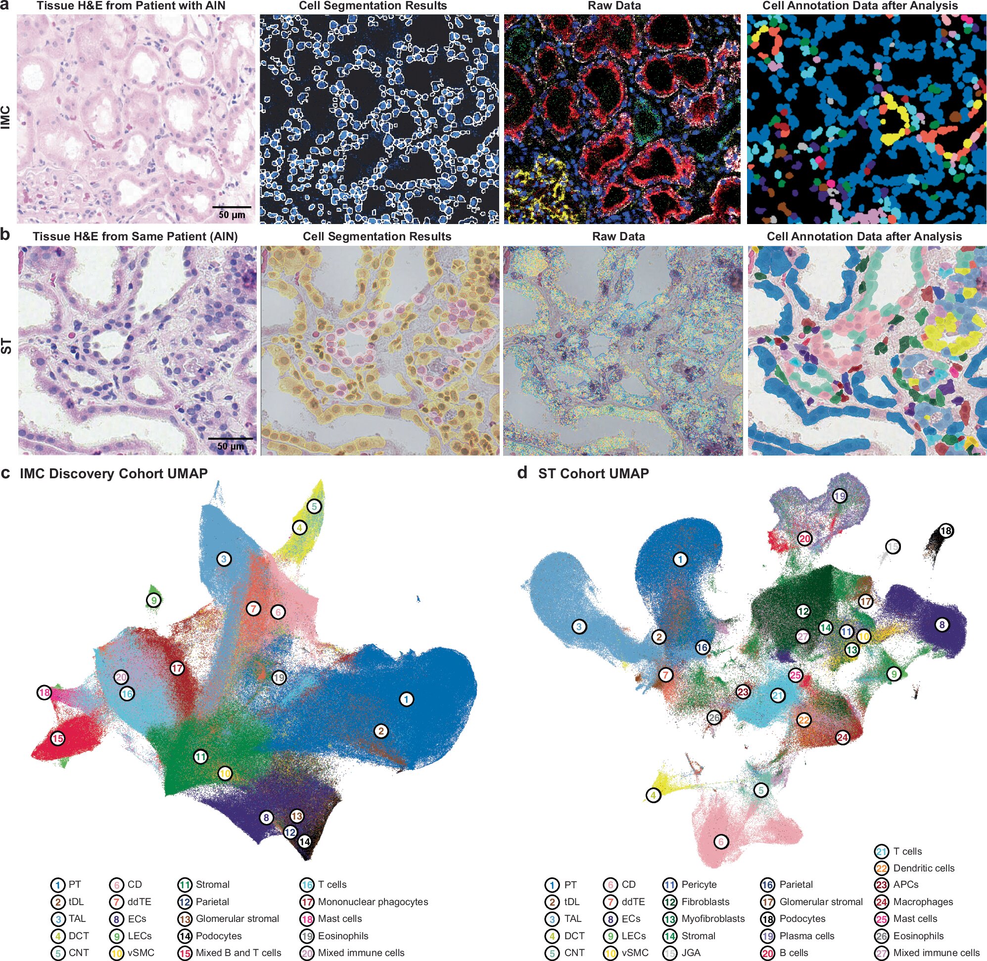
In a new study published in Nature Communications, Yale researchers used advanced cell profiling techniques to obtain the most detailed view yet of how kidney and immune cells interact in AIN.
"The standard way to measure which genes individual cells are expressing is to break the tissue apart into single cells, which destroys all of the information about how those cells were arranged and which cells were next to one another," explains the study's first author, Megan Baker, M.D., Ph.D., an instructor of medicine (nephrology) at Yale School of Medicine (YSM).
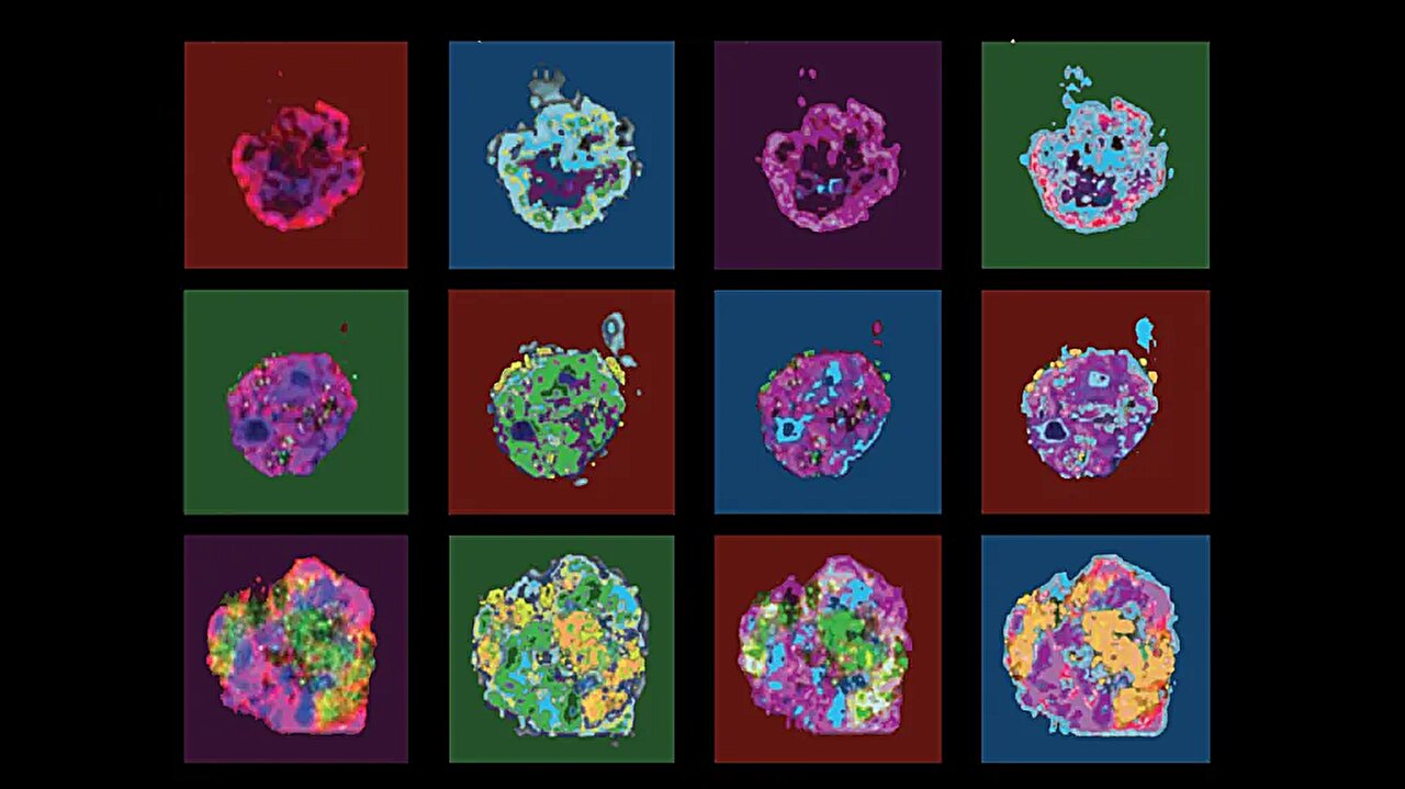
By combining technologies such as imaging mass cytometry and single-cell spatial transcriptomics, the researchers could measure gene and protein activity in individual cells while preserving each cell's location within the kidney tissue.
According to Baker, this is the first large study to map AIN at single-cell resolution while keeping each cell in place, measuring both proteins and gene expression across 106 kidney biopsies and confirming the findings in an independent group of patients at Johns Hopkins.
Source link