This article has been reviewed according to Science X's editorial process and policies. Editors have highlighted the following attributes while ensuring the content's credibility:
Avoiding accidental nerve damage is an important concern in many surgical procedures. Nerves can be difficult to distinguish from surrounding tissue, particularly in complex anatomical regions. Fluorescence-guided surgery could make them easier to see by using fluorescent agents that selectively highlight nerve tissue. But there is a practical obstacle: the fluorescence signal can be weak compared with the bright ambient light of an operating room. Surgeons may therefore need to dim or switch off the room lights during fluorescence imaging, interrupting normal surgical workflow.
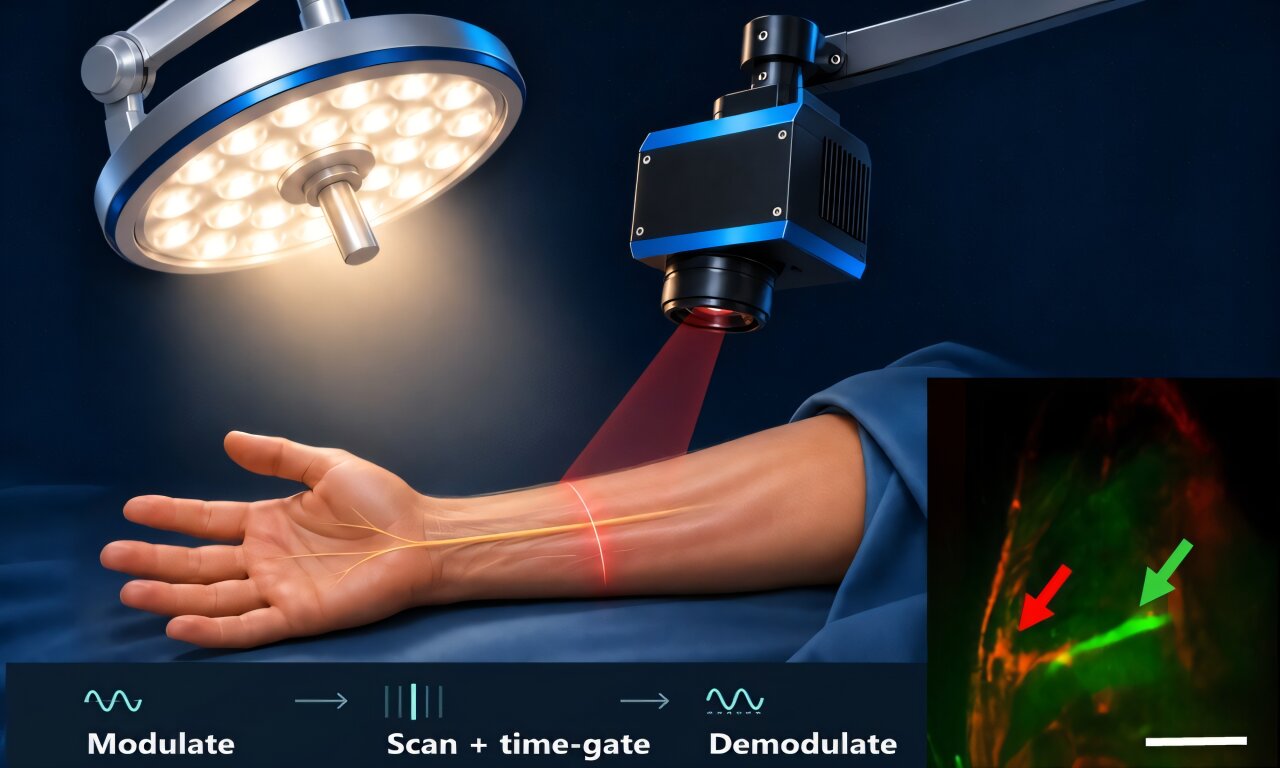
As reported in the Journal of Biomedical Optics, researchers from Physical Sciences Inc. and Dartmouth College recently developed an imaging system designed to overcome this limitation. Their approach separates fluorescence emitted by labeled nerves from the much stronger background illumination, allowing nerve signals to be visualized under standard operating-room lighting.
The problem is especially challenging for fluorescence at visible wavelengths. Light from surgical lamps can reach the camera along with the desired fluorescence, reducing contrast and potentially obscuring the structures being highlighted. Instead of trying simply to block this unwanted light, the researchers developed a way to give the fluorescence signal a recognizable spatial and temporal pattern that distinguishes it from ordinary illumination.
The system uses line-scan imaging, collecting the fluorescence signal from a narrow region at a time. This encodes the fluorescence into a specific spatial frequency and confines its detection to a short time window.
Source link