This article has been reviewed according to Science X's editorial process and policies. Editors have highlighted the following attributes while ensuring the content's credibility:
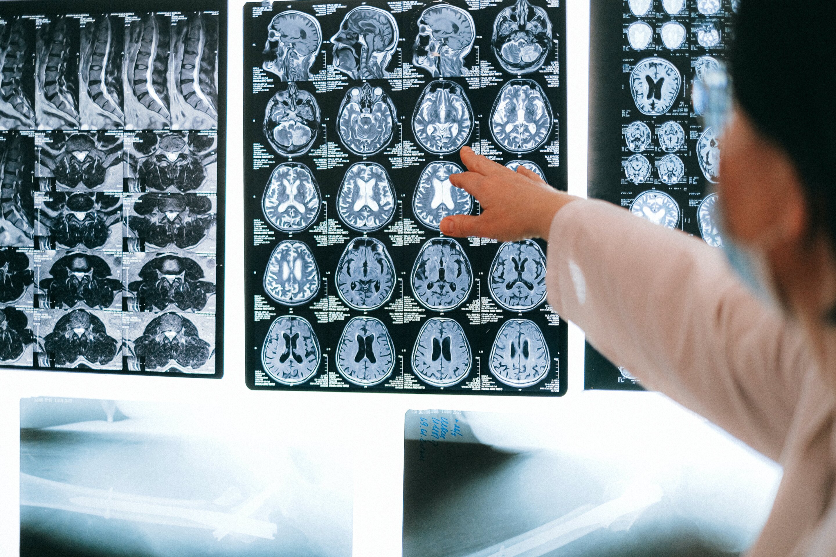
Researchers have created a large, openly available neuroimaging resource from more than 24,000 MRI scans of over 10,000 adolescents, providing a view of how the brain's wiring develops during adolescence. Using advanced, open-source image-processing methods, the team identified highly consistent patterns of development across 21 study sites, despite differences in scanner technology and image quality.
The study, led by researchers at the Perelman School of Medicine at the University of Pennsylvania with collaborators across the country, is published in Neuron.
"We showed that advanced state-of-the-art approaches to diffusion MRI can reveal aspects of adolescent brain development that conventional measures may miss," said senior author Ted Satterthwaite, M.D., the McLure II Professor of Psychiatry and Behavioral Research and director of the Penn Lifespan Informatics and Neuroimaging Center (PennLINC) at Penn Medicine.
"Despite substantial differences in image quality across tens of thousands of scans acquired by different scanners, we found remarkably consistent patterns of white matter development."
The research used data from the Adolescent Brain Cognitive Development (ABCD) Study, the largest long-term study of brain development and child health in the United States. The researchers processed more than 24,000 scans collected from over 10,000 adolescents at multiple points in time.
The team focused on diffusion MRI , a type of imaging that measures how water moves through brain tissue and can provide information about the microscopic organization of white matter—the connections that allow different parts of the brain to communicate.
Source link