This article has been reviewed according to Science X's editorial process and policies. Editors have highlighted the following attributes while ensuring the content's credibility:
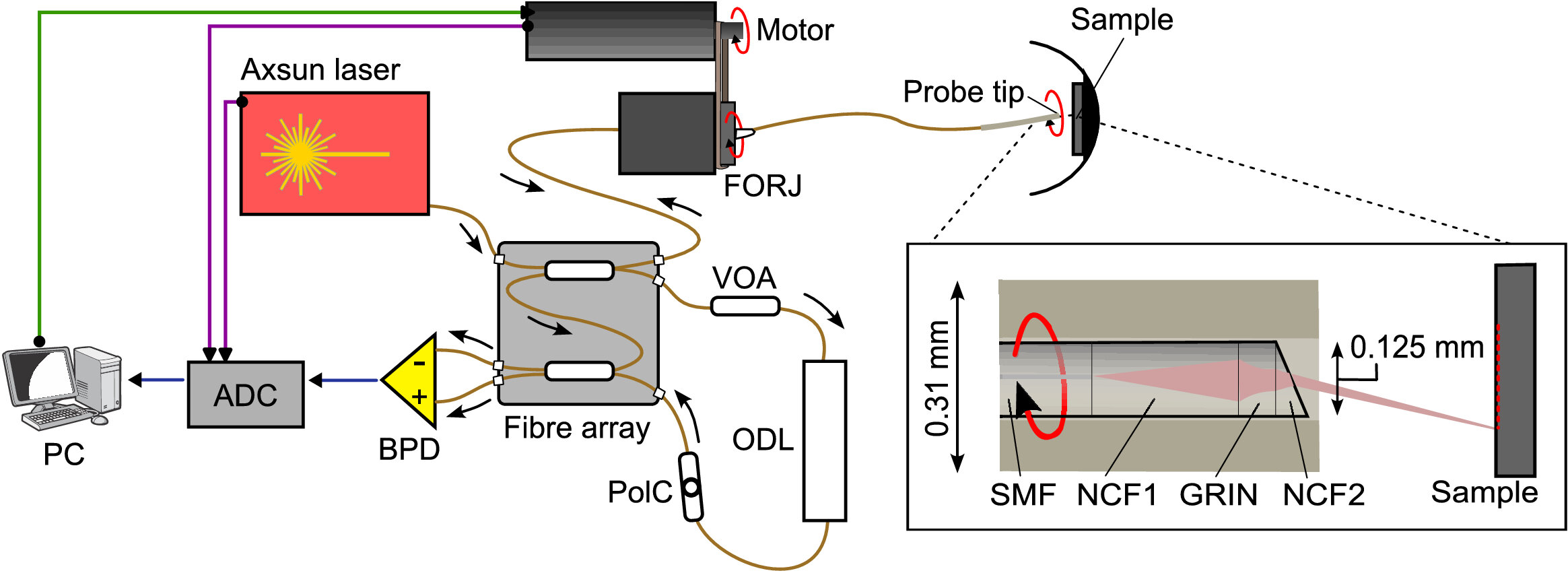
A team of physicists from the Applied Optics Group at the University of Kent has developed a new design for an ultrathin, forward-viewing endoscope. Together with partners from King's College London and Moorfields Eye Hospital, they have engineered a device that captures optical coherence tomography (OCT) images of tissue up to about 1 mm beneath its surface. The initial prototype has a diameter of just 0.31 mm, small enough for potential use in retinal surgery.
OCT is already an important tool for the diagnosis of eye disease due to its unique ability to provide high-resolution, cross-sectional images through the retina. Imaging is normally performed from outside of the eye, with images captured by rapidly scanning a low-power laser beam through the pupil. But for some surgical procedures, it may be beneficial to have a very small OCT endoscope inserted into the eye itself.
Most compact OCT endoscopes are side-viewing; when introduced into tubular structures such as blood vessels, they generate images of the vessel walls. Making an endoscope that is forward-viewing without increasing the diameter is much more difficult, but this is exactly what is needed for use inside the eye.
Dr. Radu Stancu, Dr. Michael Hughes, Dr. Manuel Marques and professor Adrian Podoleanu, all part of the School of Engineering, Mathematics and Physics, published their new endoscope design in the journal Biomedical Optics Express , where it was selected as an Editor's Pick.
Source link