This article has been reviewed according to Science X's editorial process and policies. Editors have highlighted the following attributes while ensuring the content's credibility:
University of Waterloo researchers have discovered how tiny bumps inside the eye contribute to Fuchs endothelial corneal dystrophy (FECD), an eye disease that can cause severe vision loss. With donated corneas in short supply worldwide, the finding could lead to alternative treatment strategies and reduce the need for corneal transplants in some patients.
The study, "Corneal guttata-like microtopography induces corneal endothelial cell apoptosis by modulating mechanotransduction," is published in the journal Cell Biomaterials.
"Our ultimate goal is the development of alternative treatments, which could mean fewer people will need transplants for corneal blindness due to Fuchs endothelial corneal dystrophy in the future," said Dr. Evelyn Yim, a chemical engineering professor at Waterloo and the Canada Research Chair in Nanomaterials for Regenerative Medicine.
The cornea is the clear, dome-shaped outer layer at the front of the eye that helps focus light. When it becomes cloudy, vision can become blurred or severely impaired.
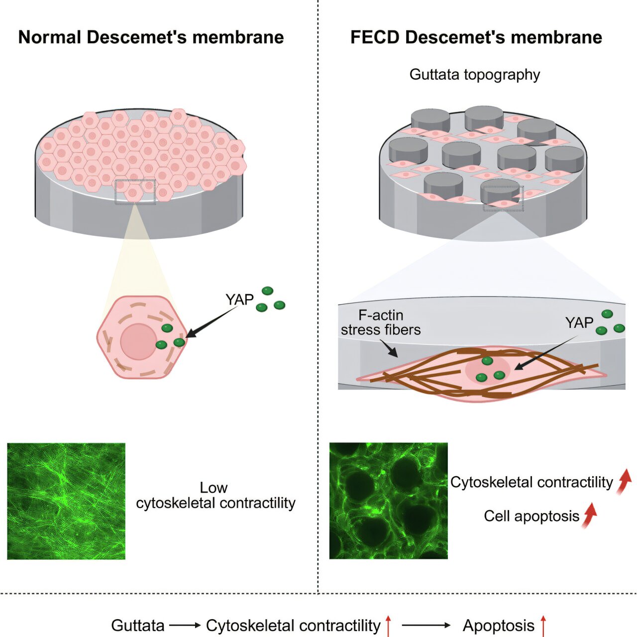
A thin layer of cells on the inside of the cornea acts like a tiny pump, constantly removing excess fluid to keep it clear. In people with FECD, these cells gradually die. As fluid builds up, the cornea swells and becomes cloudy, making it increasingly difficult to see.
For years, scientists have known that tiny bumps develop beneath these cells in people with FECD. But the bumps were largely thought to be a sign of the disease, rather than a cause of further damage.
Source link