This article has been reviewed according to Science X's editorial process and policies. Editors have highlighted the following attributes while ensuring the content's credibility:
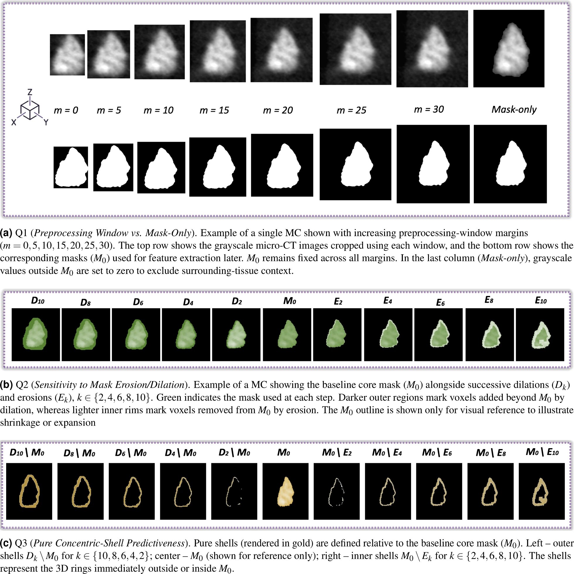
A study performed on 3,504 individual breast microcalcifications (MCs) scanned using high-resolution 3D micro-CT reveals that the surrounding tissue microenvironment provides stronger diagnostic information than the calcified core alone for distinguishing benign from malignant breast lesions. The findings are published in the journal Breast Cancer Research.
Where does the diagnostic information associated with breast MCs lie: in the calcium deposits themselves, or in the tissue immediately surrounding them? Researchers from Vrije Universiteit Brussel (VUB)-imec and UZ Brussel have addressed this question using high-resolution 3D micro-computed tomography (micro-CT) images.
The biological importance of this surrounding tissue environment is already known from histology and other research. The advance here is to isolate and compare its contribution to classification performance with that of the calcified core in micrometer-scale 3D X-ray images—for the first time, to the researchers' knowledge.
Breast MCs are well-established radiological findings in mammography and digital breast tomosynthesis. Their morphology and distribution play an important role in assessing suspicious breast lesions and determining whether further diagnostic investigation is warranted.
Traditionally, image-based assessment has focused primarily on the MCs themselves, often examining their characteristics within clusters. What has remained unclear is how much diagnostic information can be obtained from their immediate tissue surroundings in high-resolution 3D X-ray images, independently of the calcified core.
Conventional breast imaging does not provide the spatial resolution required to investigate these structures at the micrometer scale.
Source link