This article has been reviewed according to Science X's editorial process and policies. Editors have highlighted the following attributes while ensuring the content's credibility:
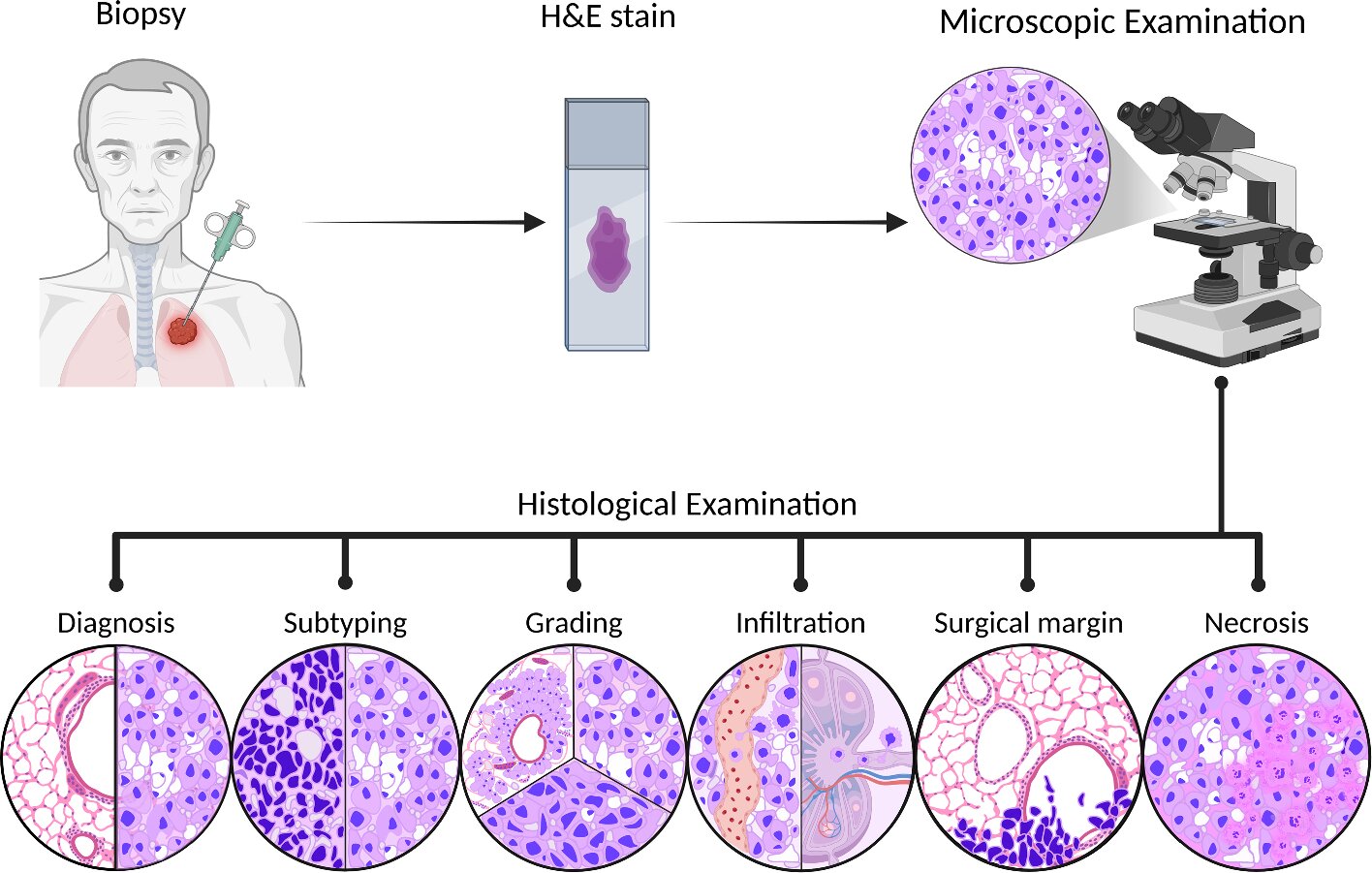
Researchers at Skoltech, the University of Sharjah in the United Arab Emirates and Tokai University in Japan, together with colleagues, have published an open-access review of computer vision in histopathology. The review examines how algorithms can identify tissue features in lung, breast, colorectal and prostate cancers.
These findings will help medical professionals and developers choose the most suitable tools for specific clinical tasks, making image analysis more accurate and reproducible. The article appears in Discover Artificial Intelligence.
Working with histological images involves a number of challenges. For example, adjacent regions of the same tumor can differ in cellular composition and tissue structure. In a digitized tissue section, pathologists assess the shape of cells and nuclei, the arrangement of tumor and immune cells, and the boundaries between a tumor and the surrounding tissue. Computer vision can help quantify these features and compare them across regions of a slide.
The features of interest depend on the clinical question. In lung cancer, algorithms help determine the tumor subtype based on tissue structure. In breast cancer, they assess tissue structure and the number of receptors—marker proteins used to select therapy—on specimens with special staining.
In colorectal cancer, one question is where immune cells are located relative to tumor foci. In prostate cancer, an important task is finding small malignant foci in a biopsy image.
As part of the review, the authors compared five tools on publicly available tissue images spanning four cancer types.
Source link