This article has been reviewed according to Science X's editorial process and policies. Editors have highlighted the following attributes while ensuring the content's credibility:
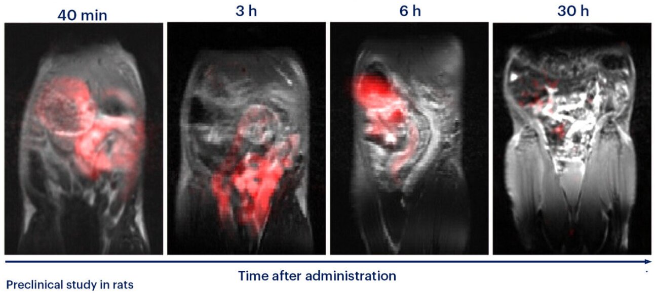
Scientists have developed a new fluorine-based tracer that can be tracked using magnetic resonance imaging (MRI) to monitor how quickly food moves through the gastrointestinal tract. The new method has so far been successfully tested in rats in a preclinical study, but it could eventually help diagnose gastrointestinal motility disorders or monitor treatment response. The study was published in Advanced Healthcare Materials.
The rate at which food moves through the stomach and intestines (gastrointestinal transit) provides important information about the health of the digestive system. Gastrointestinal transit is commonly assessed using methods that involve ionizing radiation, such as contrast X-ray imaging or scintigraphy. However, the associated radiation exposure makes these methods less suitable for frequent monitoring and limits their use in certain groups of patients, including pregnant women and children.
A team of researchers from IOCB Prague, the First Faculty of Medicine of Charles University, and Ghent University has developed a new way to monitor gastrointestinal transit using MRI, which does not involve ionizing radiation. The method relies on water-soluble, fluorinated polymer tracers that remain in the gastrointestinal tract and can be detected using fluorine-19 magnetic resonance imaging (¹⁹F MRI).
"The advantage of fluorine is that it is naturally present in the human body only in negligible amounts. This means that when we track the fluorine signal, there is virtually no background interference. We see only our tracer and can determine where it is in the gastrointestinal tract," explains Ondřej Groborz of IOCB Prague, first author of the study.
Source link