This article has been reviewed according to Science X's editorial process and policies. Editors have highlighted the following attributes while ensuring the content's credibility:
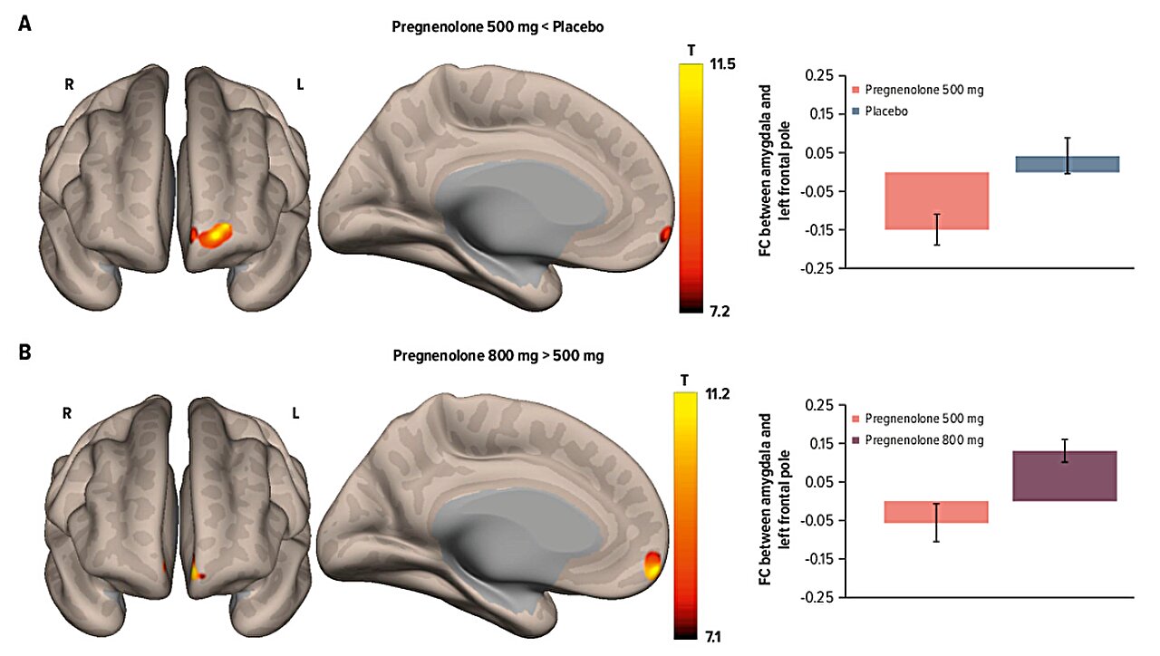
UT Southwestern Medical Center researchers and colleagues, including two at UT Dallas, have found evidence that pregnenolone—a naturally occurring steroid hormone the body produces from cholesterol—may alter patterns of communication between key brain regions involved in depression and cognitive control.
The study, conducted with the UT Dallas Center for BrainHealth and published in the Journal of Clinical Psychiatry, could potentially lead to the development of a brain-based marker of neurosteroid effects, enhancing the medical community's understanding of depression and providing insights that could inform future treatment approaches.
"Previous studies have suggested that pregnenolone could be an effective treatment for depression in people with bipolar disorder, but we lacked insight into exactly how it affects neural circuits across the brain, especially those involved in depression," said corresponding author E. Sherwood Brown, professor of psychiatry in the Peter O'Donnell Jr. Brain Institute at UT Southwestern.
"These findings bring us one step closer to developing proven neuroimaging biomarkers that would enable us to see exactly how pregnenolone changes functional connectivity between regions of the brain in people with depression—and how we might shape those changes to benefit patients."
The study followed 29 women with mild to moderate major depressive disorder in a double-blind, placebo-controlled crossover trial of pregnenolone. Participants received three seven-day treatment conditions—500 or 800 milligrams (mg) per day of pregnenolone or placebo—taken orally. Testing was performed via serum samples and functional MRI (fMRI) exams, a brain-imaging technique that enables researchers to study brain activity by measuring changes in blood flow and oxygen levels.
Source link