This article has been reviewed according to Science X's editorial process and policies. Editors have highlighted the following attributes while ensuring the content's credibility:
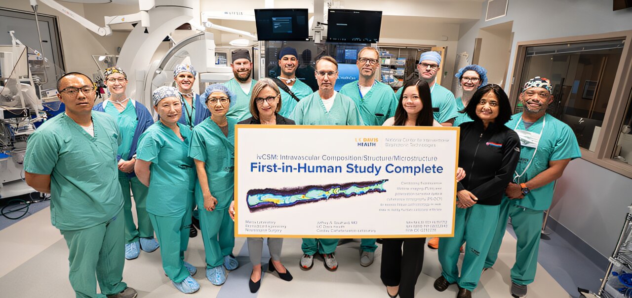
Researchers at the UC Davis National Center for Interventional Biophotonic Technologies (NCIBT) have concluded the first-in-human test of a new device that could help doctors better detect and treat atherosclerosis, the narrowing of blood vessels due to plaque buildup. The flexible catheter-based device, developed at NCIBT labs, is designed to navigate the interior walls of arteries and capture detailed information about plaque buildup in real time.
"Testing this device marks a major step toward advancing heart health. Our technology could help clinicians better understand atherosclerotic cardiovascular disease progression and personalize treatment," said Laura Marcu, founding director of NCIBT. Marcu holds the Endowed Professorship in Neurosurgical Research and is a professor in the departments of Neurological Surgery and Biomedical Engineering.
The device uses fluorescence lifetime imaging (FLIm) and polarization-sensitive optical coherence tomography (PS-OCT) to simultaneously identify the microstructure and biochemical composition of plaque in coronary arteries.
Julien Bec, who directs engineering and regulatory work in the Marcu Lab, designed and built the catheter and the imaging console.
"FLIm measures the faint natural fluorescence of the artery wall, which reflects its biochemical composition. PS-OCT measures light scattered back from the same tissue to map its microstructure," Bec explained. "The fact that we are able as an academic group to run this first-in-human study with a catheter system that was developed in a laboratory is unique.
Source link