This article has been reviewed according to Science X's editorial process and policies. Editors have highlighted the following attributes while ensuring the content's credibility:
About 60% of people diagnosed with pancreatic cancer don't qualify for surgery because their disease is too advanced. For these patients, treatment typically begins with chemotherapy, in the hope that the tumor will shrink enough to be surgically removed, which offers the best chance of long-term survival. A new clinical trial at The Ohio State University Comprehensive Cancer Center—Arthur G. James Cancer Hospital and Richard J. Solove Research Institute (OSUCCC—James) is testing a custom imaging tool designed to measure much earlier whether that chemotherapy is working, so treatment can change quickly if it is not.
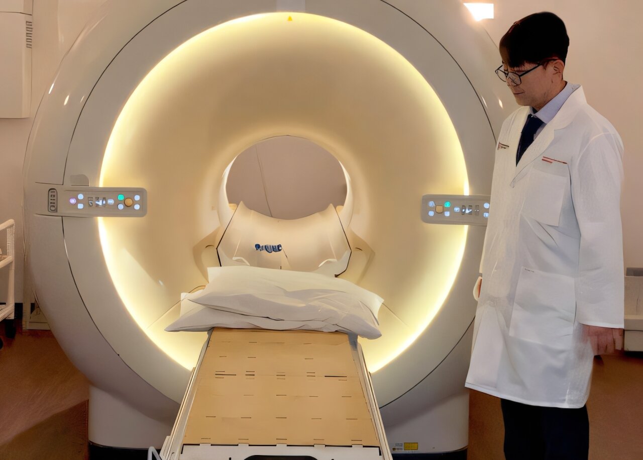
The trial will test dynamic contrast-enhanced magnetic resonance imaging (DCE-MRI) combined with standard clinical evaluation in patients with pancreatic ductal adenocarcinoma that involves arteries or veins near the pancreas but can be removed with the right treatment beforehand. DCE-MRI measures blood perfusion in the tumor, which can show disease status.
The study will enroll 50 participants and is led by Shafia Rahman, M.D., a medical oncologist at the OSUCCC—James. The tool was developed at the OSUCCC—James in partnership with Harrison Kim, Ph.D., a radiology scientist and director of the Quantitative Medical Imaging Laboratory.
"Unfortunately, the tools we have right now (CT scans) cannot give us information about how well the treatment is working to kill cancer cells earlier in the disease treatment process. This really matters in advanced pancreatic cancer because patients often experience muscle wasting and weight loss that make tolerating sustained treatment challenging," said Rahman.
Source link