This article has been reviewed according to Science X's editorial process and policies. Editors have highlighted the following attributes while ensuring the content's credibility:
For centuries, researchers and clinicians have used microscopes to study the causes and effects of disease on tissues and help make diagnoses—the practice of histopathology. While microscopes have certainly improved, scientists still rely on tissue preparation approaches that distort subtler features and entirely remove lipids and water-soluble materials. The result is an incomplete and imperfect view that cannot capture lipid data and other details that may be relevant to disease.
Researchers in the Blavatnik Institute at Harvard Medical School have developed a new imaging pipeline that overcomes these limitations and provides an unprecedented look into tissues.
The work could help researchers uncover new features associated with aging and disease and provide a basis for faster, more detailed clinical evaluation of tissues.
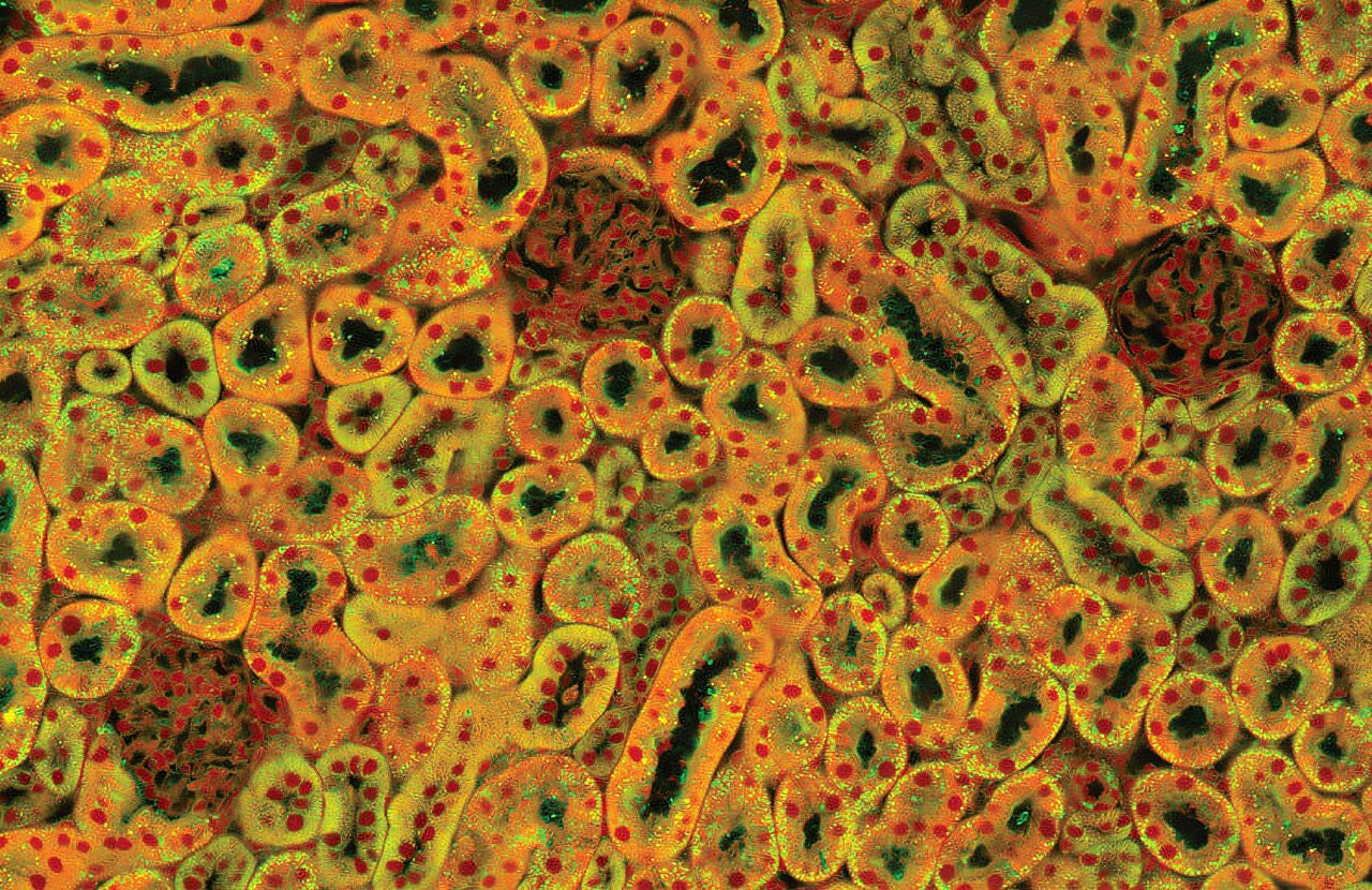
The pipeline, named NoR-AI, combines a custom microscopy method with AI-powered image analysis to capture high-quality, quantitative images that include lipids alongside proteins and water content. The technique requires little to no preprocessing of tissues and can be completed in 30 minutes, as compared to several days for traditional biopsy analysis.
In a study published in Proceedings of the National Academy of Sciences, the researchers used NoR-AI to identify previously unrecognized fine-scale changes in a mouse model of acute kidney injury.
"We're able to see into cells and tissue in ways that nobody could before," said senior author Marc Kirschner, the John Franklin Enders University Professor of Systems Biology at HMS.
Source link