Despite millions of years of evolution, the development of the human fetus remains a complex process that is vulnerable to genetic, environmental, and metabolic disruptions. These disruptions could result in developmental abnormalities, starting with one of the first organs to begin developing in the fetus that continues to develop after birth: the brain.
In a routine prenatal ultrasound, the sonographer will flag possible brain abnormalities for additional magnetic resonance imaging (MRI). But imperfect imaging can result in missed or incorrect diagnoses.
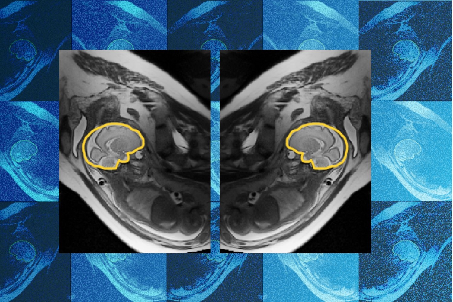
To assist with the early detection and treatment of these abnormalities, researchers at MIT, Boston Children’s Hospital, and Inria have developed and trained a deep learning model called E(3)-Pose that helps image the fetus’s head while it’s in motion to capture a high-resolution image that the radiologist can examine. A paper about the work was presented at the 2026 European Conference on Computer Vision.
“One of the problems with having the baby move is that the slices are all over the place,” says first author Ramya Muthukrishnan. Muthukrishnan is an MIT PhD student in the lab of Sunlin (1966) and Priscilla Chou Professor of Electrical Engineering and Computer Science Polina Golland, who is a principal investigator at the MIT Abdul Latif Jameel Clinic for Machine Learning in Health (Jameel Clinic) and the MIT Computer Science and Artificial Intelligence Laboratory (CSAIL).
By slices, Muthukrishnan is referring to a series of 2D images taken during a fetal MRI scan. These 2D planes stacked together effectively make up one high-resolution, 3D image of the fetus’s head.
Source link