This article has been reviewed according to Science X's editorial process and policies. Editors have highlighted the following attributes while ensuring the content's credibility:
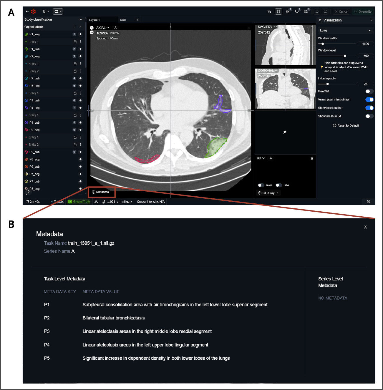
Earlier this summer, an international team led by biomedical informatics researchers at Harvard Medical School released the first public dataset that connects descriptive radiological information, such as "3 mm nodule in the lower left lobe," to precise locations in chest CT scans.
AI models trained on this dataset could analyze CT scans and communicate results more effectively with clinicians, reporting findings in descriptive language and linking those descriptions to specific pixels within three-dimensional scans.
The work, published in NEJM AI, addresses a critical gap in medical AI by making radiology findings clearer and easier to confirm, the researchers said.
"The finding becomes something a clinician can verify instead of something they have to take on faith from an AI tool," said senior author Pranav Rajpurkar, associate professor of biomedical informatics in the Blavatnik Institute at HMS.
The researchers made the dataset, ReXGroundingCT, accessible to the larger scientific community leading up to an official challenge at the International Conference on Medical Image Computing and Computer Assisted Intervention, which concluded on Sept. 27. The challenge drew 53 teams from 15 countries testing different approaches to building models that can link natural language to locations in a CT scan, a process known as grounding.
"This turns grounding from a nice idea into a benchmark people can build against," Rajpurkar said.
Source link