This article has been reviewed according to Science X's editorial process and policies. Editors have highlighted the following attributes while ensuring the content's credibility:
PSMA PET/CT-targeted biopsy detects more clinically significant prostate cancer than systematic biopsy alone in men with negative or inconclusive MRI findings but persistent clinical risk factors. According to new research published in The Journal of Nuclear Medicine, combining the two biopsy approaches is even more effective, nearly doubling the detection rate. This strategy may help physicians identify clinically significant cancers that would otherwise be difficult to detect, while potentially avoiding unnecessary procedures in appropriately selected patients.
Prostate cancer diagnosis typically involves prostate biopsy using a systematic (sampling from a predefined area) or image-guided approach. Multiparametric MRI is considered the cornerstone for detecting and localizing clinically significant prostate cancer, enabling targeted biopsy and reducing overdiagnosis of slow-growing disease. However, up to 20%–30% of clinically significant prostate cancer cases may be missed in patients with negative or inconclusive MRI findings.
"A negative or unclear MRI does not completely rule out clinically significant prostate cancer. As a result, some men may undergo repeated or extensive biopsies, while relevant tumors may still be missed," said Andrea Farolfi, M.D., a nuclear medicine physician at the Nuclear Medicine Unit, IRCCS Azienda Ospedaliero-Universitaria di Bologna, Bologna, Italy.
"Determining whether PSMA PET can refine risk stratification and guide targeted sampling for these men could have a direct impact on clinical decision-making."
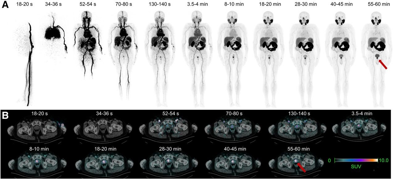
In the prospective, single-center study, 63 men with negative or inconclusive MRI scans underwent 68 Ga-PSMA-11 PET/CT followed by PET fusion-guided prostate biopsy.
Source link