This article has been reviewed according to Science X's editorial process and policies. Editors have highlighted the following attributes while ensuring the content's credibility:
Patients with early-stage lung cancer undergo surgery unless preoperative evaluation reveals poor lung function, which is predictive of poor outcomes. Imaging—like MRI or CT scans—isn't the standard of care for conventional evaluation due to issues like cost, the need for contrast and inaccuracy.
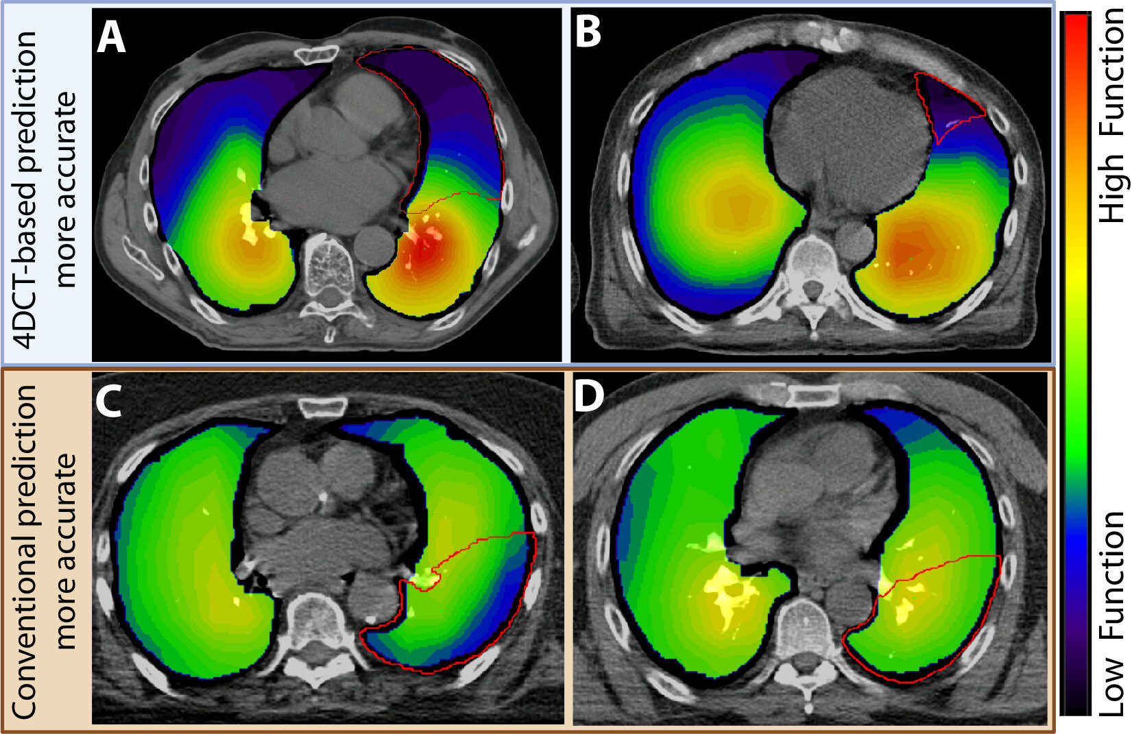
Thomas Jefferson University researchers used another imaging modality—four-dimensional computed tomography (4DCT)-ventilation—to preoperatively assess lung function in patients with early-stage lung cancer. 4DCT-ventilation is often used in radiation oncology, but this is the first time it was used in a surgical trial.
Their findings were published in the International Journal of Radiation Oncology*Biology*Physics.
"The conventional method of predicting lung function before surgery assumes that lung function is evenly distributed," says Lydia Wilson, Ph.D., the study's first author. "For patients with conditions affecting lung function, the portion you remove during surgery might not be functioning well, or it might be the only portion that's working very well, which would be detrimental to remove."
This pilot study evaluated data from 30 surgery-eligible patients with early-stage lung cancer. 4DCT-ventilation images didn't influence clinical decision-making because data analysis occurred after the trial.
While undergoing 4 DCT, patients are instructed to inhale and exhale to capture real-time lung activity. 4 DCT images create a video of the lungs moving during breathing. The researchers use image-processing techniques to convert this movie into a map showing which parts of the lung function well and which don't.
Source link