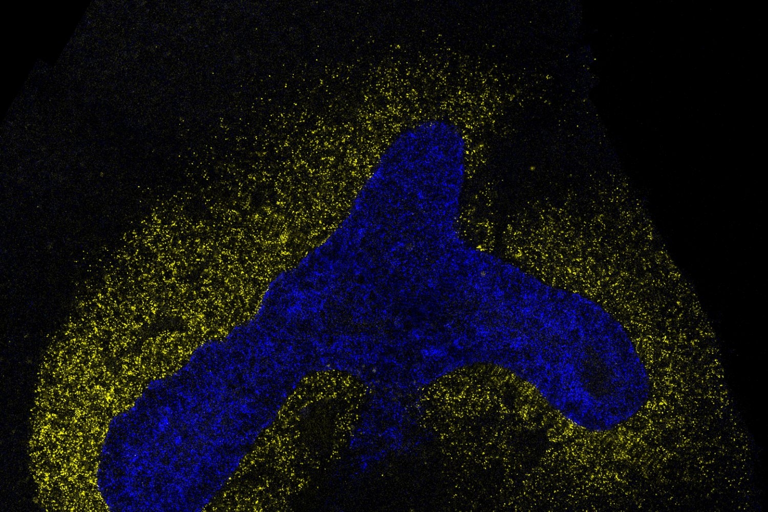
As an embryo develops from a small cluster of stem cells, those once “blank slate” cells begin to take on more specialized roles like brain, liver, or muscle cells, and organize themselves into three-dimensional structures such as tissues and organs.
The fate of each cell — what type of specialized cell it will become — depends on which genes are turned on or off in the cell. These patterns of gene activity shape the cell’s structure and function, enabling it to take on a specific role in the body.
But this decision isn’t up to individual cells. They constantly send and receive chemical signals to and from neighboring cells, which help them understand where they are, what stage of development they are in, and what they should become. These messages spread through multi-step sequences called signaling pathways, which translate external signals into specific changes in gene activity in the cell.
For researchers, being able to retrace the sequence of instructions a cell has received would offer a powerful way to understand how tissues develop and how these processes go awry in disease.
But this has been difficult to achieve because scientists have long assumed that the effects of signaling pathways vary widely across cell types, meaning they would need to map each pathway separately in each cell type — an arduous and painstaking process.
Now, in a new study led by MIT assistant professor of biology and Whitehead Institute for Biomedical Research member Pulin Li and graduate student Nicholas Hutchins, researchers have discovered that each signaling pathway leaves behind a unique “fingerprint” — a distinctive pattern of gene activity that reflects the particular signals the cell has encountered.
Source link